. 2022 Nov 28:15:1031278.
doi: 10.3389/fnmol.2022.1031278. eCollection 2022.

Cochlear resident macrophage mediates development of ribbon synapses via CX3CR1/CX3CL1 axis

Affiliations

Cochlear resident macrophage mediates development of ribbon synapses via CX3CR1/CX3CL1 axis

Xinyu Song et al. Front Mol Neurosci. .

Abstract

Cochlear ribbon synapses formed between spiral ganglion neurons and inner hair cells in postnatal mice must undergo significant morphological and functional development to reach auditory maturation. However, the mechanisms underlying cochlear ribbon synapse remodeling remain unclear. This study found that cochlear resident macrophages are essential for cochlear ribbon synapse development and maturation in mice via the CX3CR1/CX3CL1 axis. CX3CR1 expression (a macrophage surface-specific receptor) and macrophage count in the cochlea were significantly increased from postnatal day 7 then decreased from days 14 to 28. Seven-day treatment with CX3CR1 inhibitors and artificial upregulation of CX3CL1 levels in the inner ear environment using the semicircular canal injection technique were initiated on day 7, and this resulted in a significant increase in hearing threshold on day 28. Additionally, abnormalities in the morphology and number of cochlear ribbon synapses were detected on day P14, which may be associated with hearing impairment. In conclusion, macrophage regulation of cochlear ribbon synapse remodeling via the CX3CR1/CX3CL1 axis is required during hearing development and offers a new perspective on immune-related hearing loss throughout auditory development. Importantly, it could be a new treatment _target for sensorineural hearing loss.

Keywords: CX3CR1/CX3CL1; cochlear macrophage; development; hearing Loss; ribbon synapses.


Conflict of interest statement

The authors declare that the research was conducted in the absence of any commercial or financial relationships that could be construed as a potential conflict of interest.

Figures
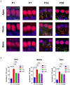
Figure 1
Figure 1
The number of synapses during postnatal mouse cochlear development is detected from P1 to P28. (A): ctbp2 dots (red) and glua2 dots (green) on immunofluorescence staining show the dynamic changes in presynaptic and postsynaptic proteins on P1, P7, P14, and p28. Scale bar = 5 μm, n = 3. The image at the bottom is an enlarged image of macrophages. (B) The number of synapses per IHCs is increased from birth to P7 until P14, after which the number of synapses are stabilized. **p < 0.01, ***p < 0.001, ****p < 0.0001.
Figure 2
Figure 2
The number of macrophages during cochlear development in postnatal mice is detected from P1 to P28. (A) Immunofluorescence staining (green) is used to detect the dynamic changes in the number of macrophages on P1, P7, P14, and p28. Scale bar = 50 μ m, n = 3. At P7, macrophages have a round shape, while they have a dendritic shape after P14. (B) The number of macrophages is increased from birth to P7 and then decreased until P14. **p < 0.01, ***p < 0.001, ****p < 0.0001.
Figure 3
Figure 3
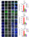
Detection of the number of CX3CR1 + macrophages during cochlear development from P1 to P28 in postnatal mice. (A) Immunofluorescence staining is used to detect the number of CX3CR1 + macrophages and identify the dynamic changes in the number of macrophages in P1. CX3CR1 (grey) colocalized with f4-80 (green). The nuclei are stained with DAPI. (B) Immunofluorescence staining is used to detect the number of CX3CR1 + macrophages and identify the dynamic changes in the number of macrophages at P7. (C) Immunofluorescence staining is used to detect the number of CX3CR1 + macrophages and identify the dynamic changes in the number of macrophages at P14. (D) Immunofluorescence staining is used to detect the number of CX3CR1 + macrophages and identify the dynamic changes in the number of macrophages at P28. Scale bar = 50 μm, n = 3. The image on the right is an enlarged image on the left. (E–G) Quantitative analysis of the number of CX3CR1 + macrophages from P1 to P28 showing that the number of CX3CR1 + macrophages from the apical to the middle to the basal turn of the cochlea is significantly higher on P7 than on P1, P14, and P28. **p < 0.01, ***p < 0.001, ****p < 0.0001.
Figure 4
Figure 4
(A) CX3CR1 expression in the cochlea of postnatal mice is determined by western blotting. A significantly stronger band is identified around P7 than in P1, P14, and P28 (n = 3). (B) Quantitative analysis of CX3CR1 expression from P1 to P28 showing that the ratio of CX3CR1 to GAPDH is significantly higher in P7 than in P1, P14, and P28. ****p < 0.0001,**p = 0.0019, ***p = 0.0003. GAPDH is used as the loading control (n = 3).
Figure 5
Figure 5
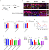
Inhibition of CX3CR1 activation in postnatal mice significantly impairs the morphological features of the ribbon synapses in cochlear IHC. (A) Mice were intraperitoneally injected with CX3CR1 inhibitor or normal saline daily starting from 7 days after birth. (B) Immunofluorescence staining of IHC ribbon synapses at P14 in each group. Presynaptic and postsynaptic proteins are co-stained; the nucleus is stained with DAPI. A magnified view of the synaptic sites is shown on the right. (C) Quantitative analysis showing that the number of synapses in the apex to the middle and bottom synapses in the cochlea is significantly higher in the CX3CR1 inhibitor-treated group than in the control group (*p < 0.05, **p < 0.01). Scale bar = 10 μm, n = 3. (D–F) Quantification of ABR threshold, amplitude, and latency of ABR wave I on P14 in control mice and CX3CR1 inhibitor mice. Significant increases in ABR thresholds at 4 and 32 kHz and click frequencies are detected in treated mice and controls. The latency of ABR wave I across frequencies (32 kHz) at P14 is longer in the treated mice than in controls. The amplitude of ABR wave I across frequencies (4 and 16 kHz) at P14 is lower in treated mice than in control mice (treatment group: n = 4, control group: n = 4; *p < 0.05, **p < 0.01).
Figure 6
Figure 6
CX3CL1 overexpression in postnatal mice significantly impairs the morphological features of ribbon synapses in cochlear IHC. (A) CX3CL1 is injected into the semicircular canal of mice 7 days after birth (B). IHC ribbon synapses in each group are subjected to immunofluorescence staining at P14. Presynaptic protein and postsynaptic protein are co-stained; the nucleus is stained with DAPI. The right side is the enlarged image of the synaptic point. (C) Quantitative analysis of synapse numbers showing that the number of synapses in the apex to the middle and bottom synapses in the cochlea is significantly higher in the CX3CL1 treatment group than in the control group (*p < 0.05, **p < 0.01). Scale bar = 10 μm, n = 3. (D–F) Quantification of ABR threshold, amplitude, and latency of ABR wave I in control mice and CX3CL1 mice on P14. Significant increases in ABR thresholds at 8 and 16 kHz and click frequencies are detected in both treated mice and control mice. The latency of ABR wave I across frequencies (16 and 32 kHz) at P14 is longer in treated mice than in control mice. The amplitude of ABR wave I across frequencies (4, 8, 16, and 32 kHz) at P14 is lower in treated mice than in control mice (treatment group: n = 4, control group: n = 4; *p < 0.05, **p < 0.01).
Figure 7
Figure 7
In the cochlea, inner hair cells convert mechanical vibrations into electrical signals via ribbon synapses at the base of cells to the auditory nerve, thereby allowing sound information to travel up the auditory pathway to the brain for processing. Cochlear spiral neurons secrete CX3CL1, which binds to CX3CR1, a specific receptor on the surface of macrophages, to phagocytose the excess weak ribbon synapses around inner hair cells during hearing development, participating in ribbon synaptic pruning.

Similar articles

Cited by

References

    1. Basilico B., Ferrucci L., Ratano P., Golia M. T., Grimaldi A., Rosito M., et al. . (2022). Microglia control glutamatergic synapses in the adult mouse hippocampus. Glia 70, 173–195. doi: 10.1002/glia.24101, PMID: - DOI - PMC - PubMed
    1. Bhuskute A., Page N. (2021). Congenital and Neonatally acquired Sensorineural hearing loss. Pediatr. Ann. 50, e292–e296. doi: 10.3928/19382359-20210629-01, PMID: - DOI - PubMed
    1. Boche D., Perry V. H., Nicoll J. A. R. (2013). Activation patterns of microglia and their identification in the human brain. Neuropathol. Appl. Neurobiol. 39, 3–18. doi: 10.1111/nan.12011, PMID: - DOI - PubMed
    1. Brites D., Fernandes A. (2015). Neuroinflammation and depression: microglia activation, extracellular microvesicles and microRNA dysregulation. Front. Cell. Neurosci. 9:476. doi: 10.3389/fncel.2015.00476 - DOI - PMC - PubMed
    1. Coate T. M., Scott M. K., Gurjar M. (2019). Current concepts in cochlear ribbon synapse formation. Synapse 73:e22087. doi: 10.1002/syn.22087, PMID: - DOI - PMC - PubMed
  NODES
twitter 2