. 2023 May;56(5):296-301.
doi: 10.5483/BMBRep.2022-0169.

Alleviation of imiquimod-induced psoriasis-like symptoms in Rorα-deficient mouse skin

Affiliations

Alleviation of imiquimod-induced psoriasis-like symptoms in Rorα-deficient mouse skin

Koog Chan Park et al. BMB Rep. 2023 May.

Abstract

Retinoic acid receptor-related orphan receptor α (RORα) plays a vital role in various physiological processes, including metabolism, cancer, circadian rhythm, cerebellar development, and inflammation. Although RORα is expressed in the skin, its role in skin physiology remains poorly elucidated. Herein, Rorα was expressed in the basal and suprabasal layers of the epidermis; however, keratinocyte-specific Rorα deletion did not impact normal epidermal formation. Under pathophysiological conditions, Rorα-deficient mice exhibited alleviated psoriasis-like symptoms, including relatively intact epidermal stratification, reduced keratinocyte hyperproliferation, and low-level expression of inflammatory cytokines in keratinocytes. Unexpectedly, the splenic population of Th17 cells was significantly lower in keratinocytespecific RORα deficient mice than in the control. Additionally, Rorα-deficiency reduced imiquimod-induced activation of nuclear factor-κB and STAT3 in keratinocytes. Therefore, we expect that RORα inhibitors act on immune cells and keratinocytes to suppress the onset and progression of psoriasis.as an adjuvant for cancer immunotherapy. [BMB Reports 2023; 56(5): 296-301].


Conflict of interest statement

CONFLICTS OF INTEREST

The authors have no conflicting interests.

Figures
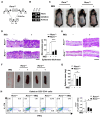
Fig. 1
Fig. 1
Evaluation of IMQ-induced psoriasis-like symptoms in skin-specific Rorα-deficient mice. (A) Rorα floxed mice were crossed with K14-Cre mice leading to keratinocyte-specific deletion of exons 4 and 5 in the Rorα gene. (B) Genotyping results for the skin-specific deletion of Rorα confirm the successful deletion of exons 4 and 5 in the Rorα gene. (C) Rorα+/+ and RorαD/D mice were topically treated with 62.5 mg of Aldara cream (containing 5% IMQ), applied daily on the shaved dorsal skin surface for six days. (D) Representative images of hematoxylin and eosin (H&E) staining of paraffin sections of the dorsal skin of each group of mice, prepared as indicated in (C) (left panel). Scale bar; 100 μm. The mean thickness of the epidermis of each group of mice was measured using H&E stained images (right panel, n = 6). Data are presented as mean ± standard error of the mean (SEM). ***P < 0.001. (E) Representative images of H&E staining of the ear skin. (F) Representative images of the spleen from each group of mice, prepared as indicated in (C). (G) The spleen mass was calculated from six independent mice for each group. Data are presented as mean ± SEM. *P < 0.05. (H) The splenic population of Th17 cells was calculated from four independent mice for each group. Data are presented as mean ± SEM. *P < 0.05. IMQ, imiquimod; RORα, Retinoic acid receptor-related orphan receptor α.
Fig. 2
Fig. 2
Analysis of epidermal stratification and barrier formation of the skin. (A-C) Skin sections were subjected to immunofluorescence analyses with antibodies against (A) Keratin 14 (K14, green) and Rorα (red), (B) K14 (green) and K10 (red), and (C) K14 (green) and loricrin (red). Scale bar = 100 μm. (D, E) Immunoblot analysis for various stratification marker proteins. Mouse primary keratinocytes (D) or HaCaT cells (E) were treated with IMQ for 24 h, and protein extracts were subjected to immunoblotting for indicated proteins. The equal protein loading was confirmed with GAPDH. (F) Skin sections were subjected to immunofluorescence analyses with antibodies against occludin, a tight junction protein. Scale bar = 100 μm. (G) The expression of occludin and claudin was analyzed as in (D). IMQ, imiquimod.
Fig. 3
Fig. 3
Assessment of IMQ-induced proliferation of wild-type and RORα-deficient human and mouse keratinocytes. (A) Skin sections were subjected to immunofluorescence analysis with an antibody against Ki67 (red), a cell proliferation marker. Scale bar = 100 μm. (B) The number of Ki67-positive keratinocyte cells was counted in four images from each independent mouse skin, expressed as a percentage of total cells. Data are shown as mean ± standard error of the mean (SEM). **P < 0.01. (C) HaCaT cells were infected with lentivirus generated from the pLentiCRISPR-E vector or pLentiCRISPR-E-RORα #1 in six-well plates. One day after viral infection, cells were selected with 1 μg/ml puromycin for 2 days. Pools of selected HaCaT cells were treated with 200 μM IMQ for 24 h and subjected to immunoblot analysis for RORα. Control represents HaCaT cells infected with lentivirus generated from the pLentiCRISPR-E vector, and RORα knockout (KO) represents the case of pLentiCRISPR-E-RORα #1. (D) HaCaT cells prepared as in (C) were seeded in 12-well plates at a density of 1 × 10⁵ cells per well, treated with IMQ for 24 h, and then EdU labeled for 24 h. EdU-positive cells indicate a red color. Scale bar = 100 μm. (E) The number of EdU-positive cells was counted in four images from each independent experiment, expressed as a percentage of total cells. Data are shown as mean ± SEM. ***P < 0.001. IMQ, imiquimod; RORα, Retinoic acid receptor-related orphan receptor α.
Fig. 4
Fig. 4
Reduced activation of IMQ-induced inflammatory signals in RORα-deficient keratinocyte cells. (A) Primary keratinocytes from the skin of newborn Rorα+/+ and RorαD/D mice were treated with 200 μM IMQ for 24 h, and cytokine levels were determined using Luminex-based multiplex assay from keratinocyte cell lysates (n = 3, three mice for each group). Data are presented as mean ± standard error of the mean (SEM). *P < 0.05; **P < 0.01, ns; not significant. (B, C) Primary keratinocytes prepared as in (A) were subjected to immunoblot analyses to evaluate the activation (phosphorylation) of NF-κB (B) and STAT3 (C). Equal protein loading was confirmed with GAPDH. (D, E) Knockout of the RORα gene was performed as described in Fig. 3C. HaCaT cells were treated with 200 μM IMQ for 24 h and subjected to immunoblot analyses to evaluate the activation (phosphorylation) of NF-κB (D) and STAT3 (E). (F) Extracts of HaCaT cells were immunoprecipitated with anti-STAT3 antibody and then blotted with antibodies indicated. (G) Control and RORα knockout HaCaT cells were treated with 200 μM IMQ for 24 h, and the expression of cytokine genes was evaluated by real-time PCR analysis. Data are presented as mean ± SEM. *P < 0.05; **P < 0.01; ***P < 0.001. IMQ, imiquimod; NF κB, nuclear factor-κB; RORα, Retinoic acid receptor-related orphan receptor α; STAT3, signal transducer and activator of transcription 3.

Similar articles

Cited by

References

    1. Lowes MA, Suarez-Farinas M, Krueger JG. Immunology of psoriasis. Annu Rev Immunol. 2014;32:227–255. doi: 10.1146/annurev-immunol-032713-120225. - DOI - PMC - PubMed
    1. Hu P, Wang M, Gao H, et al. The role of helper T cells in psoriasis. Front Immunol. 2021;12:788940. doi: 10.3389/fimmu.2021.788940.2196bd55606e40e5b9dfbfe946b234c0 - DOI - PMC - PubMed
    1. Albanesi C, Madonna S, Gisondi P, Girolomoni G. The interplay between keratinocytes and immune cells in the pathogenesis of psoriasis. Front Immunol. 2018;9:1549. doi: 10.3389/fimmu.2018.01549.dc71b21f132a47eab223f3f860f7b6db - DOI - PMC - PubMed
    1. Benhadou F, Mintoff D, Schnebert B, Thio HB. Psoriasis and microbiota: a systematic review. Diseases. 2018;6:47. doi: 10.3390/diseases6020047.5e4dbfd413c64e0f9c39780ac79d9865 - DOI - PMC - PubMed
    1. Ni X, Lai Y. Keratinocyte: a trigger or an executor of psoriasis? J Leukoc Biol. 2020;108:485–491. doi: 10.1002/JLB.5MR0120-439R. - DOI - PubMed

Grants and funding

ACKNOWLEDGEMENTS This work was supported by the Science Research Center Program (Cellular Heterogeneity Research Center, NRF-RS-2023-00207857) to KIK, and by the Basic Science Research Program (2020R111A1A01068126) to KCP, through the National Research Foundation of Korea (NRF) funded by the Ministry of Science and ICT (MSIT).
  NODES
twitter 2