. 2023 Jun 7;111(11):1795-1811.e7.
doi: 10.1016/j.neuron.2023.03.014. Epub 2023 Apr 5.

Oxytocin promotes prefrontal population activity via the PVN-PFC pathway to regulate pain

Affiliations

Oxytocin promotes prefrontal population activity via the PVN-PFC pathway to regulate pain

Yaling Liu et al. Neuron. .

Abstract

Neurons in the prefrontal cortex (PFC) can provide top-down regulation of sensory-affective experiences such as pain. Bottom-up modulation of sensory coding in the PFC, however, remains poorly understood. Here, we examined how oxytocin (OT) signaling from the hypothalamus regulates nociceptive coding in the PFC. In vivo time-lapse endoscopic calcium imaging in freely behaving rats showed that OT selectively enhanced population activity in the prelimbic PFC in response to nociceptive inputs. This population response resulted from the reduction of evoked GABAergic inhibition and manifested as elevated functional connectivity involving pain-responsive neurons. Direct inputs from OT-releasing neurons in the paraventricular nucleus (PVN) of the hypothalamus are crucial to maintaining this prefrontal nociceptive response. Activation of the prelimbic PFC by OT or direct optogenetic stimulation of oxytocinergic PVN projections reduced acute and chronic pain. These results suggest that oxytocinergic signaling in the PVN-PFC circuit constitutes a key mechanism to regulate cortical sensory processing.

Keywords: PFC; PVN; hypothalamus; nociception; oxytocin; pain; paraventricular nucleus; prefrontal cortex; prelimbic.


Conflict of interest statement

Declaration of interests The authors declare no competing interests.

Figures
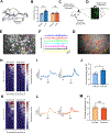
Figure 1.
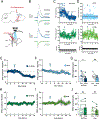
Figure 1.. Oxytocin increases population nociceptive response in the PL-PFC.
(A) Hargreaves test on a rat injected intraperitoneally (Ip) with oxytocin (OT) or saline (SAL). (B) Oxytocin prolonged the withdrawal latency. (paired t test; OT versus Baseline: ***p < 0.001; SAL vs Baseline: p = 0.5769; n = 6 animals). (C) Schematic of calcium imaging experiments. (D) Gradient-index (GRIN) lens placement and GCaMP6f expression in the PL-PFC. (E) Field of view and sample identified contours of neuronal regions of interest (ROIs). (F) Calcium activity of identified ROI traces. (G) Map of PL-PFC ROIs with contours overlaid on imaging field of view. (H) Mean Ca2+ response (z-scored) across all trials for all ROIs before and after OT. Neuronal activity is ordered from high to low responses after acute noxious pin prick (PP) stimulus. (n = 110 pre-OT, 104 post-OT ROIs). (I) Representative trace of pain-responsive neuronal ROI in response to PP before (light blue) and after (blue) OT administration. (J) Pain-responsive ROIs in the PL-PFC exhibit increased activity after OT (*p < 0.05, unpaired t test; n = 99 pre-OT, 97 post-OT ROIs). (K) Same as (H), but before and after SAL (n = 80 pre-SAL, 84 post-SAL ROIs). (L) Same as (I), but before (light orange) and after (orange) SAL. (M) Pain-responsive ROIs in the PL-PFC displayed no change in activity after SAL. (p = 0.6596, unpaired t test; n = 100 pre-SAL, 82 post-SAL ROIs). Data are represented as mean ± S.E.M. See also Figure S1, S2.
Figure 2.
Figure 2.. Oxytocin in the PL-PFC produces anti-nociceptive effects.
(A) Hargreaves test with OT delivered intracranially (Ic) to the PL-PFC. (B) Intracranial OT at 0.5 μg and 1 μg increased withdrawal latency, compared with SAL (paired t test; 1 μg OT versus baseline: ****p < 0.0001; 0.5 μg OT versus baseline: **p < 0.01, SAL versus Baseline: p = 0.2255; n = 6 animals). (C) Experimental timeline. (D) Schematic of the CPA assay, where no noxious stimulus (no prick, NP) was paired with one chamber, and noxious PP was paired with the other chamber. (E) Rats after SAL exhibited aversion to the chamber associated with PP (***p < 0.001, two-way ANOVA with repeated measures and Sidak’s multiple comparisons test; n = 6 animals). (F) Rats after OT exhibited no aversion to PP (p = 0.9998, same test as F, n = 6 animals). (G) OT decreased aversion to PP in naïve rats (**p < 0.01, paired t test; n = 6 SAL and OT animals). Data are represented as mean ± S.E.M. See also Figure S3.
Figure 3.
Figure 3.. Oxytocin in the PL-PFC reduces chronic inflammatory pain
(A) Complete Freund’s adjuvant (CFA) or saline injection to rat’s hind paw. (B) CFA decreased withdrawal threshold (****p < 0.0001, two-way ANOVA with repeated measures and Sidak’s multiple comparisons test; n = 6 animals). (C) Intracranial OT, compared SAL, increased the withdrawal threshold in CFA-treated rats (****p < 0.0001, unpaired t test; n = 6 animals). (D) Conditioned place preference (CPP) assay on tonic pain in CFA rats. Rats had SAL delivered intracranially in one chamber and OT delivered in the opposite chamber. (E) CFA-treated rats demonstrated a preference for the chamber associated with OT (**p < 0.01, n = 8 animals). (F) Control rats that received subcutaneous saline injections to paw demonstrated no preference for OT treatment (p = 0.8976, n = 8 animals). (G) CFA-treated rats exhibited a preference for the chamber associated with OT (*p < 0.05, unpaired t test; n = 8 CFA and saline animals). Data are represented as mean ± S.E.M.
Figure 4.
Figure 4.. Chemogenetic inhibition of the PVN reduces the nociceptive response in PL-PFC
(A) Viral vector and injection site of OTp-Venus in the PVN. (B) Fluorescence imaging of PVN neurons. (C) High-magnitude fluorescence imaging of PVN axons (white triangle) in the PL-PFC. (D) OTp-hM4D(Gi)-mCherry or OTp-Venus virus injection in the PVN and Gradient-index (GRIN) lens placement and GCaMP6f expression in the PL-PFC. (E) Field of view and map of example PL-PFC ROIs. (F) Representative Ca2+ activity traces of ROIs in E. (G) Mean Ca2+ response for PL-PFC ROIs imaged in sessions before and after CNO, ranked from high to low Ca2+ responses after PP. (n = 187 pre-CNO, 193 post-CNO). (H) Pain-responsive ROIs in rats injected with OTp-hM4D(Gi)-mCherry exhibited decreased Ca2+ activity after CNO (*p < 0.05, unpaired t test; n = 39 pre-CNO, 52 post-CNO). (I) Pain-responsive ROIs in rats injected with OTp-Venus showed no changes in Ca2+ activity after CNO (p = 0.9541, n = 47 pre-CNO, 58 post-CNO). Data are represented as mean ± S.E.M.
Figure 5.
Figure 5.. Optogenetic activation of the PVN axon terminals reduces synaptic inhibition in the PL-PFC
(A) Surgical (top) and recording (bottom) configurations. Blue light stimulation of ChR2-expressing PVN terminals in PL-PFC alters magnitude of post-synaptic currents evoked by local electrical stimulation (stim) recorded in whole-cell configuration (rec). (B) Example IPSC and EPSC before (gray, baseline) and after optogenetic PVN stimulation (blue, IPSC 15–20 minutes post-stimulation; green, EPSC 15–20 minutes post-stimulation). (C) Example recording showing time course of PVN stimulation (arrowhead) on IPSC amplitudes (top) and EPSC amplitudes (middle) from the same cell. Red bars, time windows for statistical analysis. Bottom, Ra was stable over the recording period; dashed lines, ±20% from average of baseline Ra. (D) Example recording showing time course of PVN stimulation with 1 µM OTA in bath; IPSCs, EPSCs, Ra were unaffected. (E,F) Summary of time course of effects of PVN stimulation (arrowhead) on IPSCs in ACSF (E, n=8 recordings) or 1 µM OTA (F, n=7 recordings) added to the bath. (G) Mean IPSC amplitudes during 5-minute baseline (Pre) and 15–20 minutes after optogenetic stimulation (Post). IPSCs were reduced by PVN stimulation in ACSF (‘No Drug’, p = 0.0128, two-way ANOVA with Bonferroni’s multiple comparisons correction, n=8 recordings), but blocking oxytocin receptors prevented this effect (‘OTA’, p=0.50, n=7 recordings). (H,I) Summary of time course of EPSCs after PVN stimulation in ACSF (left) or 1 µM OTA (right). (J) Mean EPSC amplitudes before and after PVN stimulation; no effect in either ACSF (p=0.57) or 1 µM OTA (p=0.46). Same cells as in (G). Data are represented as mean ± S.E.M.
Figure 6.
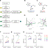
Figure 6.. Oxytocin enhances functional connectivity in pain-responsive neurons
(A) Functional connectivity analysis. We processed the raw Ca2+ imaging signal by 1) Z-scored normalization and spike deconvolution; 2) a cross-correlation matrix generation using extracted spiking activity of ROIs; 3) Monte Carlo simulation method to create 4) a matrix of statistically significant correlations between neuronal ROIs; 5) uniform subsampling of pain-responsive and non-responsive ROIs were repeated 6) for 100 iterations; 7) graph generation for each iteration. (B) Graph schematic demonstrating a node with high betweenness centrality (blue), as a central hub to multiple regions. (C) Graph schematic exhibiting a node with high degree centrality (red), as it has the most connections to other nodes. (D) An example graph from subsampled ROIs within the PL-PFC pre-OT. Green nodes: pain-responsive ROIs. Magenta nodes: ROI with the highest betweenness and degree centralities. (E) Same as (D), but post-OT. (F) Pain-responsive subpopulations demonstrated an increase in betweenness centrality (CB) post-OT (*p < 0.05, paired t test). (G) In non-responsive subpopulations, no significant change in CB was observed post-OT (p = 0.1634). (H) Pain-responsive subpopulations exhibited an increase in degree centrality (CD) after OT administration (*p < 0.05). (I) Non-responsive PL-PFC subpopulations did not exhibit a change in CD post-OT (p = 0.6738). Data are represented as mean ± S.E.M. n = 8 recordings from 5 rats. See also Figures S4, S5, and S6.
Figure 7.
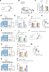
Figure 7.. Direct projection from oxytocinergic neurons in the PVN to the PL-PFC regulates acute pain
(A) Injection of OTp-ChR2-mCherry or OTp-Venus into the PVN and insertion of optic fiber into the PL-PFC. (B) Schematic of Hargreaves test. (C) Light treatment of the PL-PFC prolonged withdrawal latency in ChR2 (blue) rats, compared with control VNS (brown) rats (paired t test; ChR2 versus baseline: *p < 0.05; VNS versus baseline: p = 0.2170; n = 5 ChR2 and VNS animals). (D) CPP assay. One chamber received noxious PP paired with light treatment of the PL-PFC, and the other chamber received PP alone. (E) ChR2 rats demonstrated increased preference for treatment chamber paired with light treatment of the PL-PFC (**p < 0.01, two-way ANOVA with repeated measures and Sidak’s multiple comparisons test; n = 5 animals). (F) VNS rats exhibited no change in preference for either chamber (p = 0.9491, n = 5 animals). (G) ChR2 rats demonstrated preference for light treatment of the PL-PFC (*p < 0.05, unpaired t test; n = 5 ChR2 and n = 5 VNS animals). (H) CPP assay in the absence of noxious stimuli. One chamber was paired with blue light stimulation of the PL-PFC; the other chamber was paired with no light. (I) ChR2 rats demonstrated no preference for light treatment in the absence of noxious stimuli (p = 0.4591, n = 4 ChR2 animals). (J) Injection of OTp-ChR2-mCherry into PVN and CamKIIa-eNpHR-EYFP into PL-PFC, with optic fibers implanted in both regions. (K) CPP assay with PP. One chamber was paired with simultaneous blue light treatment of the PVN and orange light treatment of the PL-PFC; the other chamber was paired with no light treatment. (L) Rats demonstrated no preference for the chamber paired with simultaneous PVN activation and PL-PFC inhibition (p = 0.5215, n = 5 animals). (M) Rats preferred blue light activation of the PL-PFC relative to simultaneous activation of PVN and inhibition of PL-PFC (**p < 0.01, unpaired t test; n = 5 ChR2 and ChR2 + NpHR animals). (N) CPP assay with PP. In rats with both ChR2 expression in the PVN and NpHR expression in the PL-PFC, one chamber was paired with simultaneous blue light treatment of the PVN and orange light treatment of the PL-PFC, while the other chamber was paired with only blue light stimulation of the PVN. (O) Rats preferred the chamber associated with only blue light treatment of the PVN over chamber paired with simultaneous activation of PVN and inhibition of PL-PFC (*p < 0.05, n = 6 animals). Data are represented as mean ± S.E.M.
Figure 8.
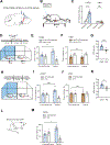
Figure 8.. Activation of axon terminals of oxytocinergic neurons from the PVN in the PL-PFC inhibits chronic pain
(A) Injection of OTp-ChR2-mCherry or OTp-Venus into the PVN and optic fiber into the PL-PFC. (B) Schematic of mechanical allodynia. (C) Light treatment of the PL-PFC increased withdrawal threshold in CFA-treated ChR2 (blue) rats, compared with VNS (control) rats. (paired t test; ChR2 versus baseline: ***p < 0.001; VNS versus baseline; p = 0.9911; n = 5 ChR2 and VNS animals). (D) CPP assay in CFA-treated rats. In one chamber, rats received noxious mechanical stimulus (6g vF) paired with light treatment of the PL-PFC. In the other chamber, rats received 6g vF alone. (E) ChR2 rats showed preference for the chamber paired with light treatment (**p < 0.01, two-way ANOVA with repeated measures and Sidak’s multiple comparisons test; n = 5 ChR2 animals). (F) VNS rats showed no preference for either chamber (p = 0.9242, n = 5 VNS animals). (G) ChR2 CFA-treated rats showed preference for optogenetic activation of the PVN-PFC pathway in the presence of 6g vF (**p < 0.01, unpaired t test; n = 5 ChR2 and VNS animals). (H) CPP assay in CFA-treated rats experiencing tonic pain. Rats received light activation of PL-PFC in one chamber and no treatment in the opposite chamber. (I) ChR2 CFA-treated rats showed preference for the chamber paired with light treatment (***p < 0.001, two-way ANOVA with repeated measures and Sidak’s multiple comparisons test; n = 5 ChR2 animals). (J) VNS CFA-treated rats showed no preference for either chamber (p = 0.9845, n = 5 VNS animals). (K) ChR2 rats experiencing tonic pain showed preference for optogenetic activation of the PVN-PFC pathway (*p < 0.05, unpaired t test; n = 5 ChR2 and VNS animals). (L) Spared-nerve injury (SNI) model. (M) ChR2 rats after SNI preferred the chamber paired with light stimulation of the PVN-PFC projection (*p < 0.05, n = 4 ChR2 animals). Data are represented as mean ± S.E.M.

Comment in

Similar articles

Cited by

References

    1. Salzman CD, and Fusi S (2010). Emotion, cognition, and mental state representation in amygdala and prefrontal cortex. Annu Rev Neurosci 33, 173–202. 10.1146/annurev.neuro.051508.135256. - DOI - PMC - PubMed
    1. Dale J, Zhou H, Zhang Q, Martinez E, Hu S, Liu K, Urien L, Chen Z, and Wang J (2018). Scaling Up Cortical Control Inhibits Pain. Cell Rep 23, 1301–1313. 10.1016/j.celrep.2018.03.139. - DOI - PMC - PubMed
    1. Hardy SG (1985). Analgesia elicited by prefrontal stimulation. Brain Res 339, 281–284. - PubMed
    1. Kiritoshi T, Ji G, and Neugebauer V (2016). Rescue of Impaired mGluR5-Driven Endocannabinoid Signaling Restores Prefrontal Cortical Output to Inhibit Pain in Arthritic Rats. J Neurosci 36, 837–850. 10.1523/JNEUROSCI.4047-15.2016. - DOI - PMC - PubMed
    1. Lee M, Manders TR, Eberle SE, Su C, D’Amour J, Yang R, Lin HY, Deisseroth K, Froemke RC, and Wang J (2015). Activation of corticostriatal circuitry relieves chronic neuropathic pain. J Neurosci 35, 5247–5259. 10.1523/JNEUROSCI.3494-14.2015. - DOI - PMC - PubMed

Publication types

  NODES
admin 2
Project 5
twitter 2