. 2023 Apr 6:14:1056154.
doi: 10.3389/fphar.2023.1056154. eCollection 2023.

Analysis of intracellular tyrosine phosphorylation in circulating neutrophils as a rapid assay for the in vivo effect of oral tyrosine kinase inhibitors

Affiliations

Analysis of intracellular tyrosine phosphorylation in circulating neutrophils as a rapid assay for the in vivo effect of oral tyrosine kinase inhibitors

Krisztina Futosi et al. Front Pharmacol. .

Abstract

Tyrosine kinases are crucial signaling components of diverse biological processes and are major therapeutic _targets in various malignancies and immune-mediated disorders. A critical step of development of novel tyrosine kinase inhibitors is the transition from the confirmation of the in vitro effects of drug candidates to the analysis of their in vivo efficacy. To facilitate this transition, we have developed a rapid in vivo assay for the analysis of the effect of oral tyrosine kinase inhibitors on basal tyrosine phosphorylation of circulating mouse neutrophils. The assay uses a single drop of peripheral blood without sacrificing the mice. Flow cytometry using intracellular staining by fluorescently labeled anti-phosphotyrosine antibodies revealed robust basal tyrosine phosphorylation in resting circulating neutrophils. This signal was abrogated by the use of isotype control antibodies or by pre-saturation of the anti-phosphotyrosine antibodies with soluble phosphotyrosine amino acids or tyrosine-phosphorylated peptides. Basal tyrosine phosphorylation was dramatically reduced in neutrophils of triple knockout mice lacking the Src-family tyrosine kinases Hck, Fgr, and Lyn. Neutrophil tyrosine phosphorylation was also abrogated by oral administration of the Abl/Src-family inhibitor dasatinib, a clinically used anti-leukemic agent. Detailed dose-response and kinetic studies revealed half-maximal reduction of neutrophil tyrosine phosphorylation by 2.9 mg/kg dasatinib, with maximal reduction observed 2 h after inhibitor administration. Taken together, our assay allows highly efficient analysis of the in vivo effect of orally administered tyrosine kinase inhibitors, and may be used as a suitable alternative to other existing approaches.

Keywords: intracellular tyrosine phosphorylation; neutrophils; rapid in vivo assay; tyrosine kinase inhibitors (TKIs); tyrosine kinases.


Conflict of interest statement

The authors declare that the research was conducted in the absence of any commercial or financial relationships that could be construed as a potential conflict of interest.

Figures

FIGURE 1
FIGURE 1
Assay overview and gating strategy. A drop of blood was stained for a neutrophil marker and for intracellular tyrosine phosphorylated proteins. A number of control and validation experiments were also performed, as indicated (A). Neutrophils were identified based on their typical forward and side scatted characteristics and positive staining for the Ly6G neutrophil marker. Tyrosine phosphorylation within those cells was then determined by intracellular flow cytometry using fluorescently labeled anti-phosphotyrosine antibodies (B). WT, wild-type; pTyr, phosphotyrosine; mAb, monoclonal antibody.
FIGURE 2
FIGURE 2
Basal tyrosine phosphorylation of circulating neutrophils. Blood was collected from untreated wild-type mice and stained with the PY20 (A–C) or the 4G10 (D–F) anti-phosphotyrosine antibodies along with isotype controls. Representative flow cytometric histograms (A, D), raw quantitative fluorescence values expressed as geometric mean fluorescence intensity (B, E) and fluorescence values normalized to those of anti-phosphotyrosine-stained samples (C, F) are shown. Bar graphs show mean and SEM from 14 (B–C) or 71 (E–F) mice from 8 (B–C) or 28 (E–F) independent experiments. MFI, geometric mean fluorescence intensity; pTyr, phosphotyrosine; Iso, isotype.
FIGURE 3
FIGURE 3
Competition by soluble phosphotyrosine or a tyrosine-phosphorylated peptide. Blood was collected from untreated wild-type mice and stained with the PY20 (A–E) or the 4G10 (F–I) anti-phosphotyrosine antibodies along with isotype controls. Where indicated, the anti-phosphotyrosine antibodies were presaturated with the indicated concentration of soluble phosphotyrosine (A, B, and E–G) or a tyrosine-phosphorylated phosphopeptide (C, D, H, and I). Representative flow cytometric histograms (A, C, F, and H) and geometric mean fluorescence intensity values normalized to those of samples stained with anti-phosphotyrosine antibodies without any presaturation with their antigens (B, D, E, G, and I) are shown. Quantitative curves and bar graphs show mean and SEM from 17 to 18 (B), 5–6 (D), 6–14 (E), 24 (G), and 6 (I) mice from 9 (B), 3 (D), 7 (E), 8 (G), and 4 (I) independent experiments. pTyr, phosphotyrosine; Iso, isotype.
FIGURE 4
FIGURE 4
Reduced basal tyrosine phosphorylation in Hck −/− Fgr −/− Lyn −/− neutrophils. Blood samples were obtained from untreated wild-type (WT) or Hck −/− Fgr −/− Lyn −/− mice and stained with the PY20 (A, B) or the 4G10 (C, D) anti-phosphotyrosine antibodies along with isotype controls. Where indicated, the anti-phosphotyrosine antibodies were presaturated with 4 mM soluble phosphotyrosine. Representative flow cytometric histograms (A, C) or geometric mean fluorescence intensity values normalized to those of WT samples stained with anti-phosphotyrosine antibodies without any presaturation with their antigens (B, D) are shown. Bar graphs show mean and SEM from 13 to 14 (B) and 10–15 (D) mice per genotype from 7 (B) and 6 (C) independent experiments. pTyr, phosphotyrosine; Iso, isotype.
FIGURE 5
FIGURE 5
Orally administered dasatinib inhibits neutrophil tyrosine phosphorylation. Wild-type mice were treated with vehicle or 50 mg/kg dasatinib by oral gavage. Blood samples were obtained 2 h later and stained with the PY20 (A, B) or the 4G10 (C, D) anti-phosphotyrosine antibodies along with isotype controls. Where indicated, the anti-phosphotyrosine antibodies were presaturated with 4 mM soluble phosphotyrosine. Representative flow cytometric histograms (A, C) or geometric mean fluorescence intensity values normalized to those of vehicle-treated samples stained with anti-phosphotyrosine antibodies without any presaturation with their antigens (B, D) are shown. Bar graphs show mean and SEM from 5 (B) or 12 (D) mice per treatment from 3 (B) or 6 (D) independent experiments. pTyr, phosphotyrosine; Iso, isotype.
FIGURE 6
FIGURE 6
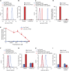
Dose-response relationship of dasatinib treatment. Wild-type mice were treated with vehicle or 1, 2, 5, 10, 20 or 50 mg/kg dasatinib by oral gavage. Blood samples were obtained 2 h later and stained with the PY20 anti-phosphotyrosine antibodies or isotype controls. Where indicated, the anti-phosphotyrosine antibodies were presaturated with 4 mM soluble phosphotyrosine. Representative flow cytometric histograms (A) or geometric mean fluorescence intensity values normalized to those of vehicle-treated samples stained with anti-phosphotyrosine antibodies without any presaturation with their antigens (B) are shown. The bar graph shows mean and SEM from 3-8 mice per treatment from at least three independent experiments. pTyr, phosphotyrosine; Iso, isotype.
FIGURE 7
FIGURE 7
Kinetic analysis of the effect of dasatinib treatment. Wild-type mice were treated with vehicle or or 50 (A), 20 (B), 10 (C), 5 mg/kg (D) dasatinib by oral gavage. Blood samples were obtained immediately before (0 time point) or 2, 5, 8 or 24 h later and stained with the PY20 anti-phosphotyrosine antibodies or isotype controls. Where indicated, the anti-phosphotyrosine antibodies were presaturated with 4 mM soluble phosphotyrosine. Geometric mean fluorescence intensity values obtained from the indicated mice and normalized to those of vehicle-treated samples stained with anti-phosphotyrosine antibodies without any presaturation with their antigens are shown. The graphs show mean and SEM from 3-8 mice per treatment from at least three independent experiments. SEM values of the normalized isotype control samples (with or without soluble phosphotyrosine pretreatment) were extremely low. pTyr, phosphotyrosine; Iso, isotype.

Similar articles

References

    1. Blaukat A. (2007). “Tyrosine kinases,” in xPharm: The comprehensive Pharmacology reference. Editors Enna S. J., Bylund D. B. (New York: Elsevier; ), 1–4.
    1. Brake K., Gumireddy A., Tiwari A., Chauhan H., Kumari D. (2017). In vivo studies for drug development via oral delivery: Challenges, animal models and techniques. Pharm. Anal. Acta 08. 10.4172/2153-2435.1000560 - DOI
    1. Breitkopf S. B., Asara J. M. (2012). Determining in vivo phosphorylation sites using mass spectrometry. Curr. Protoc. Mol. Biol. Chapter 18, 11–27. 10.1002/0471142727.mb1819s98 - DOI - PMC - PubMed
    1. Chung T. D. Y., Terry D. B., Smith L. H., Markossian S., Grossman A., Brimacombe K., et al. (Editors) (2004). “ In vitro and in vivo assessment of ADME and PK properties during lead selection and lead optimization - guidelines, benchmarks and rules of thumb,” in Assay guidance manual (Bethesda (MD): Eli Lilly and Company and the National Center for Advancing Translational Sciences; ). - PubMed
    1. Cohen P., Cross D., Janne P. A. (2021). Kinase drug discovery 20 years after imatinib: Progress and future directions. Nat. Rev. Drug Discov. 20, 551–569. 10.1038/s41573-021-00195-4 - DOI - PMC - PubMed

Grants and funding

This work was supported by the Hungarian National Research, Development and Innovation Fund (KKP-129954 and TKP2021-EGA-24 to AM) and Semmelweis University’s Scientific and Innovation Fund (STIA-2021 to AM). KF was a recipient of a János Bolyai Research Scholarship from the Hungarian Academy of Sciences. ST was supported by the National Academy of Scientist Education Program of the National Biomedical Foundation under the sponsorship of the Hungarian Ministry of Culture and Innovation.

LinkOut - more resources

  NODES
admin 6
innovation 3
twitter 2