Persistence of SARS-CoV-2 Antigens in the Nasal Mucosa of Eight Patients with Inflammatory Rhinopathy for over 80 Days following Mild COVID-19 Diagnosis
- PMID: 37112879
- PMCID: PMC10143909
- DOI: 10.3390/v15040899
Persistence of SARS-CoV-2 Antigens in the Nasal Mucosa of Eight Patients with Inflammatory Rhinopathy for over 80 Days following Mild COVID-19 Diagnosis
Abstract
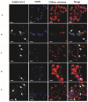
The nasal mucosa is the main gateway for entry, replication and elimination of the SARS-CoV-2 virus, the pathogen that causes severe acute respiratory syndrome (COVID-19). The presence of the virus in the epithelium causes damage to the nasal mucosa and compromises mucociliary clearance. The aim of this study was to investigate the presence of SARS-CoV-2 viral antigens in the nasal mucociliary mucosa of patients with a history of mild COVID-19 and persistent inflammatory rhinopathy. We evaluated eight adults without previous nasal diseases and with a history of COVID-19 and persistent olfactory dysfunction for more than 80 days after diagnosis of SARS-CoV-2 infection. Samples of the nasal mucosa were collected via brushing of the middle nasal concha. The detection of viral antigens was performed using immunofluorescence through confocal microscopy. Viral antigens were detected in the nasal mucosa of all patients. Persistent anosmia was observed in four patients. Our findings suggest that persistent SARS-CoV-2 antigens in the nasal mucosa of mild COVID-19 patients may lead to inflammatory rhinopathy and prolonged or relapsing anosmia. This study sheds light on the potential mechanisms underlying persistent symptoms of COVID-19 and highlights the importance of monitoring patients with persistent anosmia and nasal-related symptoms.
Keywords: COVID-19; nasal mucosa; olfactory dysfunction; rhinopathy.
Conflict of interest statement
The authors declare no conflict of interest.
Figures


Similar articles
-
COVID-19 Anosmia: High Prevalence, Plural Neuropathogenic Mechanisms, and Scarce Neurotropism of SARS-CoV-2?Viruses. 2021 Nov 4;13(11):2225. doi: 10.3390/v13112225. Viruses. 2021. PMID: 34835030 Free PMC article. Review.
-
Regeneration Profiles of Olfactory Epithelium after SARS-CoV-2 Infection in Golden Syrian Hamsters.ACS Chem Neurosci. 2021 Feb 17;12(4):589-595. doi: 10.1021/acschemneuro.0c00649. Epub 2021 Feb 1. ACS Chem Neurosci. 2021. PMID: 33522795 Free PMC article.
-
Objective evaluation of the nasal mucosal secretion in COVID-19 patients with anosmia.Ir J Med Sci. 2021 Aug;190(3):889-891. doi: 10.1007/s11845-020-02405-1. Epub 2020 Oct 19. Ir J Med Sci. 2021. PMID: 33074449 Free PMC article.
-
COVID-19-related anosmia is associated with viral persistence and inflammation in human olfactory epithelium and brain infection in hamsters.Sci Transl Med. 2021 Jun 2;13(596):eabf8396. doi: 10.1126/scitranslmed.abf8396. Epub 2021 May 3. Sci Transl Med. 2021. PMID: 33941622 Free PMC article.
-
Anosmia in COVID-19: Underlying Mechanisms and Assessment of an Olfactory Route to Brain Infection.Neuroscientist. 2021 Dec;27(6):582-603. doi: 10.1177/1073858420956905. Epub 2020 Sep 11. Neuroscientist. 2021. PMID: 32914699 Free PMC article. Review.
Cited by
-
Long COVID, the Brain, Nerves, and Cognitive Function.Neurol Int. 2023 Jul 6;15(3):821-841. doi: 10.3390/neurolint15030052. Neurol Int. 2023. PMID: 37489358 Free PMC article. Review.
-
False Positive Covid-19 Rapid Antigen Tests.N Engl J Med. 2024 May 16;390(19):1835-1836. doi: 10.1056/NEJMc2403409. N Engl J Med. 2024. PMID: 38749053 Free PMC article. No abstract available.
References
-
- Sungnak W., Huang N., Bécavin C., Berg M., Queen R., Litvinukova M., Talavera-López C., Maatz H., Reichart D., Sampaziotis F., et al. SARS-CoV-2 Entry Factors Are Highly Expressed in Nasal Epithelial Cells Together with Innate Immune Genes. Nat. Med. 2020;26:681–687. doi: 10.1038/s41591-020-0868-6. - DOI - PMC - PubMed
-
- World Health Organization Naming the Coronavirus Disease (COVID-19) and the Virus That Causes It. [(accessed on 13 February 2023)]. Available online: https://www.who.int/emergencies/diseases/novel-coronavirus-2019/technica....
-
- Chen N., Zhou M., Dong X., Qu J., Gong F., Han Y., Qiu Y., Wang J., Liu Y., Wei Y., et al. Epidemiological and Clinical Characteristics of 99 Cases of 2019 Novel Coronavirus Pneumonia in Wuhan, China: A Descriptive Study. Lancet. 2020;395:507–513. doi: 10.1016/S0140-6736(20)30211-7. - DOI - PMC - PubMed
-
- World Health Organization WHO Director—General’s Opening Remarks at the Media Briefing on COVID-19—11 March 2020. [(accessed on 13 February 2022)]. Available online: https://www.who.int/director-general/speeches/detail/who-director-genera....
Publication types
MeSH terms
Substances
Grants and funding
LinkOut - more resources
Full Text Sources
Medical
Miscellaneous

