Thymosin β4 Alleviates Autoimmune Dacryoadenitis via Suppressing Th17 Cell Response
- PMID: 37531112
- PMCID: PMC10405860
- DOI: 10.1167/iovs.64.11.3
Thymosin β4 Alleviates Autoimmune Dacryoadenitis via Suppressing Th17 Cell Response
Abstract
Purpose: We investigated the therapeutic effect of recombinant thymosin β4 (rTβ4) on rabbit autoimmune dacryoadenitis, an animal model of SS dry eye, and explore its mechanisms.
Methods: Rabbits were treated topically with rTβ4 or PBS solution after disease onset for 28 days, and clinical scores were determined by assessing tear secretion, break-up time, fluorescein, hematoxylin and eosin staining, and periodic acid-Schiff. The expression of inflammatory mediators in the lacrimal glands were measured by real-time PCR. The expression of T helper 17 (Th17) cell-related transcription factors and cytokines were detected by real-time PCR and Western blotting. The molecular mechanism underlying the effects of rTβ4 on Th17 cell responses was investigated by Western blotting.
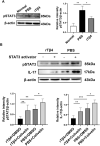
Results: Topical administration of rTβ4 after disease onset efficiently ameliorated the ocular surface inflammation and relieved the clinical symptoms. Further analysis revealed that rTβ4 treatment significantly inhibited the expression of Th17-related genes (RORC, IL-17A, IL-17F, IL-1R1, IL-23R, and granulocyte-macrophage colony-stimulating factor) and IL-17 protein in lacrimal glands, and meanwhile decreased the inflammatory mediators expression. Mechanistically, we demonstrated that rTβ4 repressed the phosphorylation of signal transducer and activator of transcription 3 (STAT3) both in vivo and in vitro. Activation of the STAT3 signal pathway by Colivelin partly reversed the suppressive effects of rTβ4 on IL-17 expression in vitro.
Conclusions: rTβ4 could alleviate ongoing autoimmune dacryoadenitis in rabbits, probably by suppressing Th17 response via partly affecting the STAT3 pathway. These data may provide a new insight into the therapeutic effect and mechanism of rTβ4 in dry eye associated with Sjögren's syndrome.
Conflict of interest statement
Disclosure:
Figures


Similar articles
-
AS101 regulates the Teff/Treg balance to alleviate rabbit autoimmune dacryoadenitis through modulating NFATc2.Exp Eye Res. 2024 Jul;244:109937. doi: 10.1016/j.exer.2024.109937. Epub 2024 May 22. Exp Eye Res. 2024. PMID: 38782179
-
PPAR-α Agonist Fenofibrate Ameliorates Sjögren Syndrome-Like Dacryoadenitis by Modulating Th1/Th17 and Treg Cell Responses in NOD Mice.Invest Ophthalmol Vis Sci. 2022 Jun 1;63(6):12. doi: 10.1167/iovs.63.6.12. Invest Ophthalmol Vis Sci. 2022. PMID: 35687344 Free PMC article.
-
Preservation of tear film integrity and inhibition of corneal injury by dexamethasone in a rabbit model of lacrimal gland inflammation-induced dry eye.J Ocul Pharmacol Ther. 2005 Apr;21(2):139-48. doi: 10.1089/jop.2005.21.139. J Ocul Pharmacol Ther. 2005. PMID: 15857280
-
Rabbit models of dry eye disease: Current understanding and unmet needs for translational research.Exp Eye Res. 2021 May;206:108538. doi: 10.1016/j.exer.2021.108538. Epub 2021 Mar 23. Exp Eye Res. 2021. PMID: 33771517 Review.
-
_targeting Th17 Effector Cytokines for the Treatment of Autoimmune Diseases.Arch Immunol Ther Exp (Warsz). 2015 Dec;63(6):405-14. doi: 10.1007/s00005-015-0362-x. Epub 2015 Sep 10. Arch Immunol Ther Exp (Warsz). 2015. PMID: 26358867 Review.
Cited by
-
Immunopeptides: immunomodulatory strategies and prospects for ocular immunity applications.Front Immunol. 2024 Jul 15;15:1406762. doi: 10.3389/fimmu.2024.1406762. eCollection 2024. Front Immunol. 2024. PMID: 39076973 Free PMC article. Review.
References
-
- Liang H, Kessal K, Rabut G, et al. .. Correlation of clinical symptoms and signs with conjunctival gene expression in primary Sjögren syndrome dry eye patients. Ocul Surf. 2019; 17: 516–525. - PubMed
-
- Yamaguchi T. Inflammatory response in dry eye. Invest Ophthalmol Vis Sci. 2018; 59: DES192–DES199. - PubMed
-
- Bjordal O, Norheim K, Rødahl E, Jonsson R, Omdal R.. Primary Sjögren's syndrome and the eye. Surv Ophthalmol. 2020; 65: 119–132. - PubMed
-
- Kojima T, Dogru M, Kawashima M, Nakamura S, Tsubota K.. Advances in the diagnosis and treatment of dry eye. Prog Retin Eye Res. 2020; 100842: 100842. - PubMed
Publication types
MeSH terms
Substances
LinkOut - more resources
Full Text Sources
Miscellaneous

