. 2023 Aug 9;13(8):1712.
doi: 10.3390/life13081712.

Characterizing 3T3-L1 MBX Adipocyte Cell Differentiation Maintained with Fatty Acids as an In Vitro Model to Study the Effects of Obesity

Affiliations

Characterizing 3T3-L1 MBX Adipocyte Cell Differentiation Maintained with Fatty Acids as an In Vitro Model to Study the Effects of Obesity

Noshin Mubtasim et al. Life (Basel). .

Abstract

The increasing prevalence of obesity has prompted intensive research into understanding its role in pathogenesis and designing appropriate treatments. To determine the signals generated from the interaction of fat cells with a _target organ, a reliable white adipocyte model in vitro is needed. Differentiated fibroblasts are the most extensively studied using in vitro cell models of white adipocytes. However, it can be argued that differentiated fibroblasts minimally recapitulate the consequences of obesity. Here, we describe 3T3-L1 MBX cells as a culture model for studying obese adipocytes and their effects. Differentiation of 3T3-L1 MBX cells was at first optimized and then maintained in the presence of fatty acids cocktail combination to induce the obese condition. Lipid accumulation and adipokine secretion profiles were analyzed. Results showed that fatty acid-maintained, differentiated 3T3-L1 MBX cells had significantly greater accumulation of lipids and significant changes in the adipokine secretions in comparison to differentiated 3T3-L1 MBX cells maintained in medium without fatty acids. To elucidate the molecular changes associated with adipogenesis and lipid accumulation profile of 3T3-L1 MBX cells, we have also explored the expression of some of the regulatory proteins related to the development and maintenance of adipocytes from the preadipocyte lineage.

Keywords: 3T3-L1 MBX; adipocyte; adipocytokine; adipogenesis; in vitro fat cell model; obesity.


Conflict of interest statement

The authors declare no conflict of interest.

Figures

Figure 1
Figure 1
Quantitation of accumulated lipids with oil-red O stain in differentiated 3T3-L1 MBX cells on Day 11 with increased concentrations of insulin and dexamethasone. (A) Cell culture images demonstrating the efficiency of differentiation in 3T3-L1 MBX cells with increased concentrations of dexamethasone (1 µM) and insulin (10 µg/mL) over 10 days post-initiation of differentiation. (B) Quantitative evaluation of the efficiency of differentiation with the dexamethasone (1 µM) and insulin (10 µg/mL) in 3T3-L1 MBX on Day 11 post-initiation of differentiation, using oil-red O stain. Each data point represents the average measure of optical density for oil-red O stain using a plate reader from 3 replicates of 3 independent experiments. The statistical significance between the treatment group and control was determined using One-way Analysis of Variance (ANOVA), and Tukey’s post hoc test was employed for multiple comparisons between group means. A p-value < 0.05 was considered statistically significant. Four asterisk (****) indicates a p-value less than 0.0001.
Figure 1
Figure 1
Quantitation of accumulated lipids with oil-red O stain in differentiated 3T3-L1 MBX cells on Day 11 with increased concentrations of insulin and dexamethasone. (A) Cell culture images demonstrating the efficiency of differentiation in 3T3-L1 MBX cells with increased concentrations of dexamethasone (1 µM) and insulin (10 µg/mL) over 10 days post-initiation of differentiation. (B) Quantitative evaluation of the efficiency of differentiation with the dexamethasone (1 µM) and insulin (10 µg/mL) in 3T3-L1 MBX on Day 11 post-initiation of differentiation, using oil-red O stain. Each data point represents the average measure of optical density for oil-red O stain using a plate reader from 3 replicates of 3 independent experiments. The statistical significance between the treatment group and control was determined using One-way Analysis of Variance (ANOVA), and Tukey’s post hoc test was employed for multiple comparisons between group means. A p-value < 0.05 was considered statistically significant. Four asterisk (****) indicates a p-value less than 0.0001.
Figure 2
Figure 2
Evaluation of 3-day cycles for differentiation induction. Three-day cycles increased the amount of accumulated lipids in 3T3-L1 MBX significantly over 2-day cycles. (A) Photomicrographs of the difference in 3 vs. 2-day cycles for inducing differentiation in 3T3-L1 MBX over 9 days. (B) Quantitative evaluation of the efficiency of 3 vs. 2-day cycles for inducing differentiation in 3T3-L1 MBX cells measured on Day 11 post-initiation, using oil-red O stain. Each data point represents the average measure of optical density of oil-red O stain using a plate reader in 3 replicates for 3 independent experiments. The statistical significance between the treatment group and the control was determined using an unpaired t-test. A p-value < 0.05 was considered statistically significant. Four asterisk (****) indicates a p-value less than 0.0001.
Figure 2
Figure 2
Evaluation of 3-day cycles for differentiation induction. Three-day cycles increased the amount of accumulated lipids in 3T3-L1 MBX significantly over 2-day cycles. (A) Photomicrographs of the difference in 3 vs. 2-day cycles for inducing differentiation in 3T3-L1 MBX over 9 days. (B) Quantitative evaluation of the efficiency of 3 vs. 2-day cycles for inducing differentiation in 3T3-L1 MBX cells measured on Day 11 post-initiation, using oil-red O stain. Each data point represents the average measure of optical density of oil-red O stain using a plate reader in 3 replicates for 3 independent experiments. The statistical significance between the treatment group and the control was determined using an unpaired t-test. A p-value < 0.05 was considered statistically significant. Four asterisk (****) indicates a p-value less than 0.0001.
Figure 3
Figure 3
Determination of the optimum non-toxic dose of individual fatty acids on differentiated 3T3-L1 MBX cells. (A) Cell viability assay determining the appropriate seeding density of 3T3-L1 MBX cell in 96-well plates for the consecutive cell viability assay against individual fatty acids for their variable mM dose range on differentiated 3T3-L1 MBX cells. mM dose range effect of (B) oleic acid, (C) linoleic acid and (D) palmitic acid on differentiated 3T3-L1 MBX cells for 24 h. Each data point represents the average measure of optical density of formazan crystal formed during MTT assay using a plate reader in 6 replicates for 3 independent experiments. The statistical significance between the treatment group and control was determined using One-way Analysis of Variance (ANOVA), and Dunnett’s multiple comparison test was employed for comparing all the dose range effects against negative control. A p-value < 0.05 was considered statistically significant. One asterisk (*) indicates p ≤ 0.05, two asterisk (**) indicates p ≤ 0.01, and three asterisk (***) indicates p ≤ 0.001. p-value > 0.05 was considered statistically nonsignificant (ns).
Figure 4
Figure 4
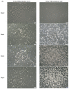
Analysis of accumulated lipids in differentiated 3T3-L1 MBX cells with and without the combination treatment of the 3 fatty acids cocktail in DMEM after the initiation of differentiation from Day 6. (A) Representative 20X images of non-oil red O stained (a) and oil-red O stained (b,c), Differentiated 3T3-L1 MBX cells cultured in DMEM in the presence and absence of fatty acids cocktail on Day 12 post-initiation of differentiation. (B) Quantitation of accumulated lipids in differentiated 3T3-L1 MBX on Days 10, 11, and 12. Each data point represents the average measure of optical density of oil-red O stain using a plate reader in 3 replicates for 3 independent experiments. The statistical significance was derived using Two-way ANOVA, and Tukey’s post hoc test was employed for multiple comparisons of group means in between. A p-value < 0.05 was considered statistically significant. Three asterisk (***) indicates p ≤ 0.001, and four asterisk (****) indicates p ≤ 0.0001.
Figure 4
Figure 4
Analysis of accumulated lipids in differentiated 3T3-L1 MBX cells with and without the combination treatment of the 3 fatty acids cocktail in DMEM after the initiation of differentiation from Day 6. (A) Representative 20X images of non-oil red O stained (a) and oil-red O stained (b,c), Differentiated 3T3-L1 MBX cells cultured in DMEM in the presence and absence of fatty acids cocktail on Day 12 post-initiation of differentiation. (B) Quantitation of accumulated lipids in differentiated 3T3-L1 MBX on Days 10, 11, and 12. Each data point represents the average measure of optical density of oil-red O stain using a plate reader in 3 replicates for 3 independent experiments. The statistical significance was derived using Two-way ANOVA, and Tukey’s post hoc test was employed for multiple comparisons of group means in between. A p-value < 0.05 was considered statistically significant. Three asterisk (***) indicates p ≤ 0.001, and four asterisk (****) indicates p ≤ 0.0001.
Figure 5
Figure 5
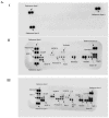
Adipocyte secretions from differentiated 3T3-L1 MBX cells on Day 11 with and without the combination treatment of 3 kinds of fatty acids in DMEM after the initiation of differentiation from Day 6. (A) The dot blots indicate different concentrations of adipokines detected on nitrocellulose membrane after treatment with supernatants of (I) DMEM media without FBS, which has been used for conditioning cells (II) 3T3-L1 MBX cells from Day 11 treated with FBS-free DMEM in the absence of fatty acids cocktail (III) 3T3-L1 MBX cells from Day 11 treated with FBS-free DMEM in the presence of fatty acids cocktail. (B) Bar graphs of adipokines with the observable changes in adipokine secretions from differentiated 3T3-L1 MBX cells cultured in the presence or absence of fatty acids cocktail. Each data point represents the mean value of the secretome in 2 technical replicates for 3 independent experiments. (C) Fold change expression of secreted adipokines from differentiated 3T3-L1 MBX cells on Day 11 treated with fatty acids cocktail in respect to differentiated 3T3-L1 MBX cells on Day 11 treated without fatty acids. The statistical significance between the treatment group and the control was determined using a paired t-test. A p-value < 0.05 was considered statistically significant. One asterisk (*) indicates p ≤ 0.05, two asterisk (**) indicates p ≤ 0.01, and three asterisk (***) indicates p ≤ 0.001. p-value > 0.05 was considered statistically nonsignificant (ns).
Figure 5
Figure 5
Adipocyte secretions from differentiated 3T3-L1 MBX cells on Day 11 with and without the combination treatment of 3 kinds of fatty acids in DMEM after the initiation of differentiation from Day 6. (A) The dot blots indicate different concentrations of adipokines detected on nitrocellulose membrane after treatment with supernatants of (I) DMEM media without FBS, which has been used for conditioning cells (II) 3T3-L1 MBX cells from Day 11 treated with FBS-free DMEM in the absence of fatty acids cocktail (III) 3T3-L1 MBX cells from Day 11 treated with FBS-free DMEM in the presence of fatty acids cocktail. (B) Bar graphs of adipokines with the observable changes in adipokine secretions from differentiated 3T3-L1 MBX cells cultured in the presence or absence of fatty acids cocktail. Each data point represents the mean value of the secretome in 2 technical replicates for 3 independent experiments. (C) Fold change expression of secreted adipokines from differentiated 3T3-L1 MBX cells on Day 11 treated with fatty acids cocktail in respect to differentiated 3T3-L1 MBX cells on Day 11 treated without fatty acids. The statistical significance between the treatment group and the control was determined using a paired t-test. A p-value < 0.05 was considered statistically significant. One asterisk (*) indicates p ≤ 0.05, two asterisk (**) indicates p ≤ 0.01, and three asterisk (***) indicates p ≤ 0.001. p-value > 0.05 was considered statistically nonsignificant (ns).
Figure 6
Figure 6
The expression of adipogenesis-related proteins in 3T3-L1 MBX cells from its journey from preadipocyte to adipocyte lineage w/o the treatment of fatty acids cocktail. (A) Immunoblotting image and (B) protein expression level of adipogenesis-related protein in 3T3-L1 MBX cells on Day 0, Day 3, Day 6, and Day 11 (with and without the fatty acids cocktail treatment). Each data point represents the average measure of densitometric quantification of protein expression for 3 independent biological replicates for each Day of protein extract. The statistical significance was derived using One-way ANOVA, and Dunnett’s multiple comparison test was employed for comparing the expression of regulatory proteins in differentiated days (Day 3, 6, and 11) to Day 0 when the 3T3-L1 MBX cells were in non-differentiated condition. A p-value < 0.05 was considered statistically significant. One asterisk (*) indicates p ≤ 0.05, two asterisk (**) indicates p ≤ 0.01, and three asterisk (***) indicates p ≤ 0.001. p-value greater than 0.05 was considered statistically non-significant (ns). Here: FA: fatty acid; PPAR-γ: Peroxisome Proliferator- Activated Receptor gamma; CBP: CREB binding protein; GCN5L2: General Control of Amino Acid Synthesis Yeast Homolog Like 2; AMPK-α: AMP-Activated Protein Kinase, RXR-α: cis-Retinoic Acid Receptor.
Figure 6
Figure 6
The expression of adipogenesis-related proteins in 3T3-L1 MBX cells from its journey from preadipocyte to adipocyte lineage w/o the treatment of fatty acids cocktail. (A) Immunoblotting image and (B) protein expression level of adipogenesis-related protein in 3T3-L1 MBX cells on Day 0, Day 3, Day 6, and Day 11 (with and without the fatty acids cocktail treatment). Each data point represents the average measure of densitometric quantification of protein expression for 3 independent biological replicates for each Day of protein extract. The statistical significance was derived using One-way ANOVA, and Dunnett’s multiple comparison test was employed for comparing the expression of regulatory proteins in differentiated days (Day 3, 6, and 11) to Day 0 when the 3T3-L1 MBX cells were in non-differentiated condition. A p-value < 0.05 was considered statistically significant. One asterisk (*) indicates p ≤ 0.05, two asterisk (**) indicates p ≤ 0.01, and three asterisk (***) indicates p ≤ 0.001. p-value greater than 0.05 was considered statistically non-significant (ns). Here: FA: fatty acid; PPAR-γ: Peroxisome Proliferator- Activated Receptor gamma; CBP: CREB binding protein; GCN5L2: General Control of Amino Acid Synthesis Yeast Homolog Like 2; AMPK-α: AMP-Activated Protein Kinase, RXR-α: cis-Retinoic Acid Receptor.

Similar articles

Cited by

References

    1. Blüher M. Obesity: Global epidemiology and pathogenesis. Nat. Rev. Endocrinol. 2019;15:288–298. doi: 10.1038/s41574-019-0176-8. - DOI - PubMed
    1. Coelho M., Oliveira T., Fernandes R. Biochemistry of adipose tissue: An endocrine organ. Arch. Med. Sci. 2013;9:191–200. doi: 10.5114/aoms.2013.33181. - DOI - PMC - PubMed
    1. Poddar M., Chetty Y., Chetty V.T. How does obesity affect the endocrine system? A narrative review. Clin. Obes. 2017;7:136–144. doi: 10.1111/cob.12184. - DOI - PubMed
    1. Dufau J., Shen J.X., Couchet M., De Castro Barbosa T., Mejhert N., Massier L., Griseti E., Mouisel E., Amri E.Z., Lauschke V.M., et al. In vitro and ex vivo models of adipocytes. Am. J. Physiol. Cell Physiol. 2021;320:C822–C841. doi: 10.1152/ajpcell.00519.2020. - DOI - PubMed
    1. Mubtasim N., Moustaid-Moussa N., Gollahon L. The Complex Biology of the Obesity-Induced, Metastasis-Promoting Tumor Microenvironment in Breast Cancer. Int. J. Mol. Sci. 2022;23:2480. doi: 10.3390/ijms23052480. - DOI - PMC - PubMed

LinkOut - more resources

  NODES
Association 1
twitter 2