. 2023 Oct 5;11(10):1563.
doi: 10.3390/vaccines11101563.

Usp22 Deficiency Leads to Downregulation of PD-L1 and Pathological Activation of CD8+ T Cells and Causes Immunopathology in Response to Acute LCMV Infection

Affiliations

Usp22 Deficiency Leads to Downregulation of PD-L1 and Pathological Activation of CD8+ T Cells and Causes Immunopathology in Response to Acute LCMV Infection

Justa Friebus-Kardash et al. Vaccines (Basel). .

Abstract

Ubiquitin-specific peptidase 22 (Usp22) cleaves ubiquitin moieties from numerous proteins, including histone H2B and transcription factors. Recently, it was reported that Usp22 acts as a negative regulator of interferon-dependent responses. In the current study, we investigated the role of Usp22 deficiency in acute viral infection with lymphocytic choriomeningitis virus (LCMV). We found that the lack of Usp22 on bone marrow-derived cells (Usp22fl/fl Vav1-Cre mice) reduced the induction of type I and II interferons. A limited type I interferon response did not influence virus replication. However, restricted expression of PD-L1 led to increased frequencies of functional virus-specific CD8+ T cells and rapid death of Usp22-deficient mice. CD8+ T cell depletion experiments revealed that accelerated CD8+ T cells were responsible for enhanced lethality in Usp22 deficient mice. In conclusion, we found that the lack of Usp22 generated a pathological CD8+ T cell response, which gave rise to severe disease in mice.

Keywords: LCMV; PD-L1 downregulation; activation of CD8+ T cells; liver failure; ubiquitin-specific peptidase 22 (Usp22).


Conflict of interest statement

The authors declare no conflict of interest.

Figures

Figure 1
Figure 1
Usp22 deficiency is associated with the reduced production of type I and II interferons and with a slight increase in virus titers at the early stage of acute LCMV infection. (AC) Usp22 deficient and WT mice were infected intravenously with LCMV WE (high dose, 2 × 10⁵ PFU per mouse) and examined at the indicated time points. (A) Serum levels of interferons α, β, and γ were measured in the serum by multiplex cytokine assay in Usp22 KO and WT mice on days 0, 1, 4, 7, and 9 after infection (n = 6). (B) LCMV titers from the spleen were measured by plaque assay on day 2 after infection (n = 6). (C) Viral titers were determined by plaque assay in the liver, spleen, lung, and kidney on day 9 after infection (n = 6). Data are shown as mean ± SEM and pooled from 2 to 3 independent experiments. *, p = 0.05; (AC) unpaired two-tailed Student’s t-test.
Figure 2
Figure 2
Conditional deletion of Usp22 in mice leads to downregulation of PD-L1 expression on antigen-presenting cells after acute LCMV infection. (AD) We administered 2 × 10⁵ PFU of the LCMV WE intravenously to each mouse on day 0. (A,B) We measured PD-L1 expression on CD11b+Ly6C+ Ly6G monocytes. The percentages of CD11b+Ly6C+ Ly6G cells positive for PD-L1 (A) and the expression of PD-L1 on CD11b+Ly6C+ Ly6G cells (B) in the peripheral blood (n = 6) and spleen (n = 9) were determined by flow cytometry at the indicated time points. (C,D) Decreased expression of PD-L1 in the spleen (C) and liver (D) tissue of Usp22 KO and WT mice obtained on day 3 after infection and detected by immunofluorescence analysis. Scale bar = 100 μm; original magnification ×10; one representative of six is shown. Data are presented as mean ± SEM and are pooled from 2 to 3 independent experiments. **, p = 0.01; ***, p = 0.001; (A,B) unpaired two-tailed Student’s t-test.
Figure 3
Figure 3
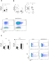
LCMV infection in Usp22-deficient mice results in an inflammatory response characterized by the activation of virus-specific CD8+ T cells. (AC) Usp22-deficient mice and WT mice were infected intravenously with LCMV WE (2 × 10⁵ PFU per mouse) on day 0 and examined at the indicated time points. (A) LCMV-specific GP33 positive CD8+ T cells were assessed in the peripheral blood by tetramer staining and by flow cytometry on day 7 after infection (n = 10). (B) Numbers of CD8+ T cells in liver and spleen tissue obtained on day 9 after infection (n = 6) were determined by flow cytometry. (C) Intracellular IFN-γ production by CD8+ T cells derived from spleen and liver tissue of Usp22 KO mice and WT control mice (n = 6) was determined by flow cytometry after re-stimulation with LCMV specific peptides (gp33). Data from two or three independent experiments with consistent results are shown. *, p = 0.05; **, p = 0.01; (AC) unpaired two-tailed Student’s t-test.
Figure 4
Figure 4
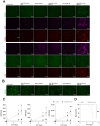
After acute LCMV infection, a deficiency in Usp22 leads to infiltration of the liver by monocytes and neutrophil granulocytes and to subsequent acute liver failure with a lethal outcome. (A) Immunofluorescence of liver sections from Usp22 KO and WT mice performed on days 3 and 9 after infection, stained for CD11b-, CD11c-, Ly6G-, and Ly6C-positive cells, CD8+ T cells, and F4/80+ macrophages. Scale bar = 100 μm; one representative of six is shown. Fluorescent microscopy images were captured at 10× magnification with a Keyence BZ-9000E microscope. (B) Three and nine days later, immunofluorescence analysis was conducted for CD169+ macrophages in snap-frozen spleen tissue from Usp22 KO and WT mice. Scale bar = 100 μm; one representative of six is shown. Original magnification ×10. (C,D) We injected 2 × 10⁵ PFU LCMV WE intravenously into each mouse on day 0. (C) Liver enzyme levels were monitored for the indicated period after LCMV infection (n = 11). (D) Survival analysis during acute LCMV infection, comparing Usp22 KO mice (n = 11) and WT mice (n = 11). Data are combined from 2 to 3 independent experiments with consistent results. *, p = 0.05; **, p = 0.01; (C) unpaired two-tailed Student’s t-test; (D) log-rank (Mantel–Cox).
Figure 5
Figure 5
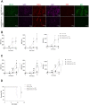
Although the depletion of CD8+ T cells rescues Usp22 deficient mice from acute immunopathology after LCMV infection, the depletion of neutrophil granulocytes and monocytes only delays the development of liver failure in Usp22 KO mice. (AD) Before infection with LCMV WE (2 × 10⁵ PFU per mouse), Usp22 deficient mice and WT mice were treated intraperitoneally with 100 μg of the monoclonal anti-CD8a antibody (A) or with 500 μg of the anti-Gr-1 antibody (C) per mouse on day −1, day 0, and then every second day. PBS served as a control. Intravenous infection with LCMV WE (2 × 10⁵ PFU per mouse) was performed on day 0. (A) Immunofluorescence staining of snap-frozen liver sections was performed on day 9 after depletion of CD8+ T cells with subsequent infection. Scale bar = 100 μm; one representative of six is shown. Fluorescence microscopy images were captured at 10× magnification with a Keyence BZ-9000E microscope. (B,C) Liver enzyme levels were determined at the indicated time points after infection and before administration of monoclonal anti-CD8a antibody (B) or anti-Gr-1 antibody (C) (n = 6). (D) Survival rates of Usp22 KO and WT mice after LCMV infection and subsequent treatment with CD8+ T cell depleting antibody (n = 6). Results from 2 independent experiments are pooled. Data are shown as mean ± SEM. *, p = 0.05; **, p = 0.01; ***, p = 0.001; (B,C) unpaired two-tailed Student’s t-test; (D) log-rank (Mantel–Cox).

Similar articles

Cited by

References

    1. Rehermann B., Nascimbeni M. Immunology of hepatitis B virus and hepatitis C virus infection. Nat. Rev. Immunol. 2005;5:215–222. doi: 10.1038/nri1573. - DOI - PubMed
    1. Smatti M.K., Cyprian F.S., Nasrallah G.K., Al Thani A.A., Almishal R.O., Yassine H.M. Viruses and autoimmunity: A review on the potential interaction and molecular mechanisms. Viruses. 2019;11:762. doi: 10.3390/v11080762. - DOI - PMC - PubMed
    1. Knight J.S., Caricchio R., Casanova J.L., Combes A.J., Diamond B., Fox S.E., Hanauer D.A., James J.A., Kanthi Y., Ladd V., et al. The intersection of COVID-19 and autoimmunity. J. Clin. Investig. 2021;131:e154886. doi: 10.1172/JCI154886. - DOI - PMC - PubMed
    1. Seo Y.J., Hahm B. Type I interferon modulates the battle of host immune system against viruses. Adv. Appl. Microbiol. 2010;73:83–101. - PMC - PubMed
    1. Teijaro J.R., Ng C., Lee A.M., Sullivan B.M., Sheehan K.C., Welch M., Schreiber R.D., Carlos de la Torre J., Oldstone M.B. Persistent LCMV infection is controlled by blockade of type I interferon signaling. Science. 2013;340:207–211. doi: 10.1126/science.1235214. - DOI - PMC - PubMed
  NODES
admin 2
twitter 2