. 2023 Dec;63(6):140.
doi: 10.3892/ijo.2023.5588. Epub 2023 Nov 3.

MEK/ERK and PI3K/AKT pathway inhibitors affect the transformation of myelodysplastic syndrome into acute myeloid leukemia via H3K27me3 methylases and de‑methylases

Affiliations

MEK/ERK and PI3K/AKT pathway inhibitors affect the transformation of myelodysplastic syndrome into acute myeloid leukemia via H3K27me3 methylases and de‑methylases

Zhuanzhen Zheng et al. Int J Oncol. 2023 Dec.

Abstract

The transformation of myelodysplastic syndrome (MDS) into acute myeloid leukemia (AML) poses a significant clinical challenge. The trimethylation of H3 on lysine 27 (H3K27me3) methylase and de‑methylase pathway is involved in the regulation of MDS progression. The present study investigated the functional mechanisms of the MEK/ERK and PI3K/AKT pathways in the MDS‑to‑AML transformation. MDS‑AML mouse and SKM‑1 cell models were first established and this was followed by treatment with the MEK/ERK pathway inhibitor, U0126, the PI3K/AKT pathway inhibitor, Ly294002, or their combination. H3K27me3 methylase, enhancer of zeste homolog (EZH)1, EZH2, demethylase Jumonji domain‑containing protein‑3 (JMJD3) and ubiquitously transcribed tetratricopeptide repeat on chromosome X (UTX) and H3K27me3 protein levels were determined using western blot analysis. Cell viability, cycle distribution and proliferation were assessed using CCK‑8, flow cytometry, EdU and colony formation assays. The ERK and AKT phosphorylation levels in clinical samples and established models were determined, and SKM‑1 cell behaviors were assessed. The levels of H3K27me3 methylases and de‑methylases and distal‑less homeobox 5 (DLX5) were measured. The results revealed that the ERK and AKT phosphorylation levels were elevated in patients with MDS and MDS‑AML, and in mouse models. Treatment with U0126, a MEK/ERK pathway inhibitor, and Ly294002, a PI3K/AKT pathway inhibitor, effectively suppressed ERK and AKT phosphorylation in mice with MDS‑AML. It was observed that mice with MDS treated with U0126/Ly294002 exhibited reduced transformation to AML, delayed disease transformation and increased survival rates. Treatment of the SKM‑1 cells with U0126/Ly294002 led to a decrease in cell viability and proliferation, and to an increase in cell cycle arrest by suppressing ERK/PI3K phosphorylation. Moreover, treatment with U0126/Ly294002 downregulated EZH2/EZH1 expression, and upregulated JMJD3/UTX expression. The effects of U0126/Ly294002 were nullified when EZH2/EZH1 was overexpressed or when JMJD3/UTX was inhibited in the SKM‑1 cells. Treatment with U0126/Ly294002 also resulted in a decreased H3K27me3 protein level and H3K27me3 level in the DLX5 promoter region, leading to an increased DLX5 expression. Overall, the findings of the present study suggest that U0126/Ly294002 participates in MDS‑AML transformation by modulating the levels of H3K27me3 methylases and de‑methylases, and regulating DLX5 transcription and expression.

Keywords: H3K27me3; Ly294002; MEK/ERK; PI3K/AKT; SKM‑1 cells; U0126; acute myeloid leukemia; distal‑less homeobox 5; myelodysplastic syndrome.


Conflict of interest statement

The authors declare that they have no competing interests.

Figures

Figure 1
Figure 1
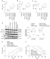
Abnormal activation of the MEK/ERK and PI3K/AKT pathways in patients with MDS and MDS-AML. Western blot analysis was performed to determine the phosphorylation levels of ERK and AKT in bone marrow samples of patients with MDS and MDS-AML and cancer-free individuals. Multi-group comparisons were performed using one-way ANOVA, followed by Tukey's multiple comparisons test. ***P<0.001. MDS, myelodysplastic syndrome; AML, acute myeloid leukemia.
Figure 2
Figure 2
MEK/ERK and PI3K/AKT pathway inhibitors attenuate the MDS-AML transformation in NHD13 mice. (A-C) The numbers of WBCs, RBCs and PLTs in C57B/L6 mice or NHD13 mice in the different groups were counted using a full-automatic blood cell analyzer. (D) The protein levels of ERK, p-ERK, AKT and p-AKT in the peripheral blood of C57B/L6 mice or NHD13 mice in the different groups were determined using western blot analysis. (E) The numbers of NHD13 mice in which MDS transformed to AML within 14 months (420 days) and the transformation time. (F) The survival rates of NHD13 mice were analyzed using Kaplan-Meier survival curves. (A-D) n=6; multi-group comparisons were performed using one-way ANOVA, followed by Tukey's multiple comparisons test. *P<0.05, **P<0.01 and ***P<0.001. (E and F) n=12; comparisons between groups were performed using the log-rank test. p-, phosphorylated; MDS, myelodysplastic syndrome; AML, acute myeloid leukemia; WBC, white blood cells; RBC, red blood cell; PLT, platelet.
Figure 3
Figure 3
MEK/ERK and PI3K/AKT pathway inhibitors regulate the SKM-1 cell cycle and proliferation. (A) Western blot analysis was performed to determine the protein levels of ERK, p-ERK, AKT and p-AKT in SKM-1 cells. (B) SKM-1 cell viability was assessed using CCK-8 assay. (C) SKM-1 cell cycle progression was evaluated using flow cytometry. (D and E) SKM-1 cell proliferation was assessed using EdU and colony formation assays. Each cell experiment was repeated three times. The results are expressed as the mean ± standard deviation. Multi-group comparisons were performed using one-way ANOVA, followed by Tukey's multiple comparisons test. *P<0.05 and ***P<0.001.
Figure 4
Figure 4
MEK/ERK and PI3K/AKT pathway inhibitors modulate the expression levels of H3K27me3 methylases and de-methylases. Western blot analysis was performed to determine the protein levels of H3K27me3 methylases EZH1, EZH2, and de-methylases JMJD3 and UTX, and H3K27me3 in SKM-1 cells. Each cell experiment was repeated three times. The results are expressed as the mean ± standard deviation. Multi-group comparisons were performed using one-way ANOVA, followed by Tukey's multiple comparisons test. *P<0.05 and ***P<0.001. H3K27me3, trimethylation of H3 on lysine 27; EZH, enhancer of zeste homolog; JMJD3, demethylase Jumonji domain-containing protein-3; UTX, ubiquitously transcribed tetratricopeptide repeat on chromosome X.
Figure 5
Figure 5
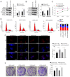
EZH2 overexpression or JMJD3 knockdown partially reverses the suppressive effects of MEK/ERK pathway inhibitors on SKM-1 cells. SKM-1 cells were transfected with EZH2 overexpression plasmid oe-EZH2 or JMJD3 interference plasmid si-JMJD3 for 48 h and then treated with the MEK/ERK inhibitor, U0126 (5 µM) for 48 h. (A and B) The protein levels of EZH2 or JMJD3 and H3K27me3 were measured using western blot analysis. (C) SKM-1 cell viability was assessed using CCK-8 assay. (D) SKM-1 cell cycle distribution was analyzed using flow cytometry. (E and F) SKM-1 cell proliferation was evaluated using EdU and colony formation assays. Each cell experiment was repeated three times. The results are expressed as the mean ± standard deviation. Pairwise comparison were analyzed using an independent t-test. *P<0.05. EZH2, enhancer of zeste homolog 2; JMJD3, demethylase Jumonji domain-containing protein-3; H3K27me3, trimethylation of H3 on lysine 27.
Figure 6
Figure 6
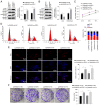
EZH1 overexpression or UTX knockdown partially annuls the suppressive effects of PI3K/AKT pathway inhibitors on SKM-1 cells. SKM-1 cells were transfected with EZH1 overexpression plasmid oe-EZH1 or UTX interference plasmid si-UTX for 48 h and then treated with the PI3K/AKT inhibitor, Ly294002 (5 µM) for 48 h. (A and B) The protein levels of EZH1 or UTX and H3K27me3 were measured using western blot analysis. (C) SKM-1 cell viability was assessed using CCK-8 assay. (D) SKM-1 cell cycle distribution was analyzed using flow cytometry. (E and F) SKM-1 cell proliferation was evaluated using EdU and colony formation assays. Each cell experiment was repeated three times. The results are expressed as the mean ± standard deviation. Pairwise comparison was analyzed using an independent t-test. *P<0.05. EZH1, enhancer of zeste homolog 1; H3K27me3, trimethylation of H3 on lysine 27; UTX, ubiquitously transcribed tetratricopeptide repeat on chromosome X.
Figure 7
Figure 7
MEK/ERK and PI3K/AKT pathway inhibitors facilitate DLX5 transcription and expression by downregulating H3K27me3. (A) The level of DLX5 promoter region binding to H3K27me3 was examined using ChIP assay, and the graph shows RT-qPCR analysis of the binding content of the DLX5 promoter in DNA IP after ChIP, which is expressed as the content relative to control DNA input; the columns revealed the detection results of DLX5 ChIP primer 1, and the results were validated using DLX5 ChIP primer 2. The detection results of the two primers were similar. (B and C) DLX5 mRNA and protein levels in SKM-1 cells were measured using RT-qPCR and western blot analysis. Each cell experiment was repeated three times. The results are expressed as the mean ± standard deviation. Multi-group comparisons were performed using one-way ANOVA, followed by Tukey's multiple comparisons test. *P<0.05 and ***P<0.001. DLX5, distal-less homeobox 5; H3K27me3, trimethylation of H3 on lysine 27; RT-qPCR, reverse transcription-quantitative PCR.
Figure 8
Figure 8
Schematic diagram of the role of the MEK/ERK and PI3K/AKT pathways in MDS-AML. The MEK/ERK and PI3K/AKT pathway inhibitors, U0126 and Ly294002, respectively, regulated te H3K27me3 level by mediating the levels of EZH2/JMJD3 and EZH1/UTX, thus affecting the transcription and expression of DLX5, and ultimately participating in the regulation of MDS and AML progression. MDS, myelodysplastic syndrome; AML, acute myeloid leukemia; EZH, enhancer of zeste homolog; H3K27me3, trimethylation of H3 on lysine 27; UTX, ubiquitously transcribed tetratricopeptide repeat on chromosome X; JMJD3, demethylase Jumonji domain-containing protein-3; JMJD3, demethylase Jumonji domain-containing protein-3.

Similar articles

Cited by

References

    1. Hasserjian RP. Myelodysplastic syndrome updated. Pathobiology. 2019;86:7–13. - PubMed
    1. Haferlach T. The molecular pathology of myelodysplastic syndrome. Pathobiology. 2019;86:24–29. - PubMed
    1. Yan X, Lai B, Zhou X, Yang S, Ge Q, Zhou M, Shi C, Xu Z, Ouyang G. The differential expression of CD47 may be related to the pathogenesis from myelodysplastic syndromes to acute myeloid leukemia. Front Oncol. 2022;12:872999. - PMC - PubMed
    1. Menssen AJ, Walter MJ. Genetics of progression from MDS to secondary leukemia. Blood. 2020;136:50–60. - PMC - PubMed
    1. Rankin EB, Narla A, Park JK, Lin S, Sakamoto KM. Biology of the bone marrow microenvironment and myelodysplastic syndromes. Mol Genet Metab. 2015;116:24–28. - PMC - PubMed

MeSH terms

Grants and funding

The present study was funded by The Shanxi Province Applied Basic Research: Natural Science Foundation (grant no. 201801D121330), which aims to investigate the association between molecular biology abnormalities based on epigenetic modifiers and mRNA precursor splicing factors and the prognosis of myelodysplastic syndromes.
  NODES
Association 1
twitter 2