. 2024 Feb 28;52(4):1896-1908.
doi: 10.1093/nar/gkad1217.

Conversion of the CG specific M.MpeI DNA methyltransferase into an enzyme predominantly methylating CCA and CCC sites

Affiliations

Conversion of the CG specific M.MpeI DNA methyltransferase into an enzyme predominantly methylating CCA and CCC sites

Pál Albert et al. Nucleic Acids Res. .

Abstract

We used structure guided mutagenesis and directed enzyme evolution to alter the specificity of the CG specific bacterial DNA (cytosine-5) methyltransferase M.MpeI. Methylation specificity of the M.MpeI variants was characterized by digestions with methylation sensitive restriction enzymes and by measuring incorporation of tritiated methyl groups into double-stranded oligonucleotides containing single CC, CG, CA or CT sites. Site specific mutagenesis steps designed to disrupt the specific contacts between the enzyme and the non-substrate base pair of the _target sequence (5'-CG/5'-CG) yielded M.MpeI variants with varying levels of CG specific and increasing levels of CA and CC specific MTase activity. Subsequent random mutagenesis of the _target recognizing domain coupled with selection for non-CG specific methylation yielded a variant, which predominantly methylates CC dinucleotides, has very low activity on CG and CA sites, and no activity on CT sites. This M.MpeI variant contains a one amino acid deletion (ΔA323) and three substitutions (N324G, R326G and E305N) in the _target recognition domain. The mutant enzyme has very strong preference for A and C in the 3' flanking position making it a CCA and CCC specific DNA methyltransferase.


Figures

Graphical Abstract
Graphical Abstract
Figure 1.
Figure 1.
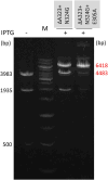
Testing the sequence specificity of wild-type M.MpeI by digesting the plasmid pET28-MMpeI with Alw44I. (A) Positions of the Alw44I cleavage sites, sizes of the fragments obtained after complete digestion (in bp, blue), and the CN sites (in red) created by the 3′-C of the Alw44I recognition sequence and the 3′-flanking nucleotide (see above the gel). (B) Fragment patterns of Alw44I digested plasmids purified from uninduced and IPTG induced cultures. Size of the protected fragment is shown in red. M, GeneRuler 1 kb DNA Ladder.
Figure 2.
Figure 2.
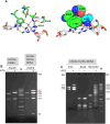
Selected features of sequence specific DNA recognition by M.MpeI as revealed by X-ray crystallography (15). (A) Amino acid sequence of the recognition loops. (B) Schematic representation of sequence specific interactions between the enzyme and the _target site. Carbon 5 of the _target cytosine carries a fluorine atom, whereas the cytosine in the non-substrate strand of the palindromic recognition site is methylated in the C5 position (15).
Figure 3.
Figure 3.
Methylation specificity of M.MpeI(A323G+E305A) and M.MpeI(A323G+S325G+E305A). (A) Interactions maintaining the stability of the H-bond between Ala323 and the guanine-O6 of the CG substrate site (15). Left panel, hydrogen bonds between the Ser325 and the oxygen of the peptide bond between Gly322 and Ala323; Right panel, pocket formed by Recognition Loop 1 residues holding the methyl group of Ala323. (B) Digestion of pET28-MMpeI(A323G+E305A) and pET28-MMpeI(A323G+S325G+E305A) with Alw44I. (C) Digestion of pET28-MMpeI(A323G+S325G+E305A) with XmiI, BsuRI and Bsh1236I. Fragment sizes in red letters indicate undigested fragments. For interpretation of the digestion patterns see the plasmid maps in Figure 1 and Supplementary Figure S1. M, GeneRuler 1 kb DNA Ladder.
Figure 4.
Figure 4.
Alw44I digestion of pET28-MMpeI expressing the indicated M.MpeI variants. Sizes of the protected fragments are shown in red. For interpretation of the fragment pattern, see Figure 1. M, GeneRuler 1 kb DNA Ladder.
Figure 5.
Figure 5.
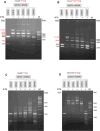
Digestion of pET28-MMpeI(ΔA323+N324G+E305X) plasmids with Alw44I, XmiI, BsuRI and Bsh1236I. On panels A and B sizes of the protected fragments are shown in red. For interpretation of the Alw44I and XmiI fragment pattern, see Figure 1 and Supplementary Figure S1, respectively. The plasmids contain 23 BsuRI and 35 Bsh1236I sites. The substrate site whose methylation status can be tested by digestion with the respective restriction enzyme is shown in red above the gels. M, GeneRuler 1 kb DNA Ladder.
Figure 6.
Figure 6.
Testing the methylation specificity of M.MpeI(ΔA323+N324G+R326G+E305N). (A) Positions of Eco47I cleavage sites, the sizes of the fragments obtained after complete digestion (in bp, blue), and the CN sites (in red) created by the 3′-C of the Eco47I recognition sequence GGWCC and the 3′-flanking nucleotide. The unique XmiI site (not shown on the map) is flanked by adenine on both sides. (B) Digestion of pOB-MMpeI(ΔA323+N324G+R326G+E305N) with methylation sensitive restriction enzymes. Plasmid preparations marked by + signs were purified from cells induced with arabinose for five hours. The methylation specificity tested with the particular enzyme is shown in red above the enzyme's name. M, GeneRuler 1 kb DNA Ladder. The digestion patterns were reproduced in three independent experiments.
Figure 7.
Figure 7.
Testing the specificity of M.MpeI(ΔA323+N324G+R326G+E305N) by in vitro MTase assay using purified enzyme, [methyl-3H] labeled SAM and double stranded oligonucleotide substrates. In the substrate oligonucleotides (CG, AK702-AK703; CA, AK704-AK705; CT, AK706-AK707; CC, AK708-AK709) the substrate site was in AXXA context. (A) [methyl-3H] radioactivity incorporated by M.MpeI(ΔA323+N324G+R326G+E305N) in 30 min reactions. Average values of two independent experiments. (B and C) Time course of methyl transfer by wild-type M.MpeI and M.MpeI(ΔA323+N324G+R326G+E305N) into the CG, CA and CC duplexes. Average values of three independent experiments. The concentration of wild-type and mutant M.MpeI was 5 and 350 nM, respectively. Error bars: standard error of the mean.
Figure 8.
Figure 8.
Effects of the flanking nucleotides on the CC-specific activity of M.MpeI(ΔA323+N324G+R326G+E305N). In vitro MTase reactions using purified enzyme, [methyl-3H] labeled SAM and double-stranded oligonucleotide substrates. [methyl-3H] radioactivity incorporated in 30 min reactions. Average values of three measurements. Two nucleotides preceding and following the CC substrate site are shown. To highlight the flanking nucleotides, the cc core substrate site is shown with lower case letters. Sequences of the substrate oligonucleotides are shown in Supplementary Tables S2 and S6. Error bars: standard error of the mean.
Figure 9.
Figure 9.
Steady-state kinetic analysis of methyl transfer into double stranded oligonucleotides containing the indicated substrate sites. The reactions contained 5 nM wild-type or 350 nM mutant M.MpeI and 2 μM 5′-ACGA/5′-Tm5CGT (AK702–AK703) and 5′-TCCA/5′-TGGA (AK923-AK924) duplex, respectively. Error bars: standard error of the mean.

Similar articles

References

    1. Bochtler M., Fernandes H. DNA adenine methylation in eukaryotes: enzymatic mark or a form of DNA damage?. Bioessays. 2021; 43:2000243. - PubMed
    1. Rodriguez F., Yushenova I.A., DiCorpo D., Arkhipova I.R. Bacterial N4-methylcytosine as an epigenetic mark in eukaryotic DNA. Nat. Commun. 2022; 13:1072. - PMC - PubMed
    1. Jurkowska R.Z., and Jeltsch A. (2022) Mechanisms and biological roles of DNA methyltransferases and DNA methylation: from past achievements to future challenges. In: Jeltsch A., Jurkowska R.Z. (eds.) DNA Methyltr ansfer ases - Role and Function. Advances in Experimental Medicine and Biology. Vol. 1389, Springer, Cham, pp. 1–19. - PubMed
    1. Pósfai J., Bhagwat A.S., Pósfai G., Roberts R.J. Predictive motifs derived from cytosine methyltransferases. Nucleic Acids Res. 1989; 17:2421–2435. - PMC - PubMed
    1. Goll M.G., Bestor T.H. Eukaryotic cytosine methyltransferases. Annu. Rev. Biochem. 2005; 74:481–514. - PubMed

MeSH terms

  NODES
twitter 2