. 2024 Jan 19;12(1):226.
doi: 10.3390/biomedicines12010226.

A Brain-Protective Sterol from Soft Coral Inhibits Lipopolysaccharide-Induced Matrix Metalloproteinase-9-Mediated Astrocytic Migration

Affiliations

A Brain-Protective Sterol from Soft Coral Inhibits Lipopolysaccharide-Induced Matrix Metalloproteinase-9-Mediated Astrocytic Migration

Tsong-Hai Lee et al. Biomedicines. .

Abstract

Matrix metalloproteinases (MMPs), which are proteolytic enzymes, promote blood-brain barrier (BBB) disruption, leading to neuronal damage and neuroinflammation. Among them, MMP-9 upregulation serves as an inflammatory biomarker in the central nervous system (CNS). Currently, the development of marine organism-derived bioactive compounds or metabolites as anti-inflammatory drugs has received considerable attention. The 9,11-secosteroid, 3β,11-dihydroxy-9,11-secogorgost-5-en-9-one (4p3f), is a novel sterol compound extracted from the soft coral Sinularia leptoclado with potential anti-inflammatory activity. However, the effect of and potential for brain protection of 4p3f on brain astrocytes remain unclear. Herein, we used rat brain astrocytes (RBAs) to investigate the effects and signaling mechanisms of 4p3f on lipopolysaccharide (LPS)-induced MMP-9 expression via zymographic, quantitative reverse transcription-polymerase chain reaction (qRT-PCR), Western blot, immunofluorescence staining, promoter-reporter, and cell migration analyses. We first found that 4p3f blocked LPS-induced MMP-9 expression in RBAs. Next, we demonstrated that LPS induced MMP-9 expression via the activation of ERK1/2, p38 MAPK, and JNK1/2, which is linked to the STAT3-mediated NF-κB signaling pathway. Finally, 4p3f effectively inhibited LPS-induced upregulation of MMP-9-triggered RBA cell migration. These data suggest that a novel sterol from soft coral, 4p3f, may have anti-inflammatory and brain-protective effects by attenuating these signaling pathways of MMP-9-mediated events in brain astrocytes. Accordingly, the soft coral-derived sterol 4p3f may emerge as a potential candidate for drug development or as a natural compound with neuroprotective properties.

Keywords: anti-inflammation; brain astrocytes; cell migration; lipopolysaccharide; matrix metalloproteinase-9; soft coral.


Conflict of interest statement

The authors declare no conflicts of interest.

Figures

Figure 1
Figure 1
The effect of a soft coral-derived sterol extract on LPS-induced MMP-9 expression in brain astrocytes. (A,B) Cells were treated with LPS (10 μg/mL) for the indicated time intervals. (A) For MMP-9 expression, the conditioned media were collected and analyzed via gelatin zymography. And the glyceraldehyde-3-phosphate dehydrogenase (GAPDH) level of cell lysates (as an internal control) was assayed by means of Western blot analysis. (B) Total RNA was prepared and then MMP-9 mRNA expression was analyzed via real-time RT-PCR. (C,D) Cells were pretreated with 4p3f (1 μM) for 1 h and then incubated with LPS for the indicated times. (C) The conditioned media were collected and analyzed for MMP-9 expression via gelatin zymography. (D) MMP-9 mRNA expression was analyzed by means of real-time RT-PCR. Data are expressed as mean ± SEM (n = 3). * p < 0.05 and ** p < 0.01, as compared with the untreated control; # p < 0.05, as compared with LPS-treated cells only. The images represent one of three individual experiments.
Figure 2
Figure 2
4p3f attenuates LPS-induced MMP-9 expression by blocking MAPK signaling pathways. Cells were pretreated with U0126 (0.1 μM), SB202190 (1 μM), or SP600125 (0.1 μM) for 1 h and then incubated with LPS (10 μg/mL) for the indicated time intervals. (A) For MMP-9 expression, the conditioned media were collected and analyzed via gelatin zymography. The GAPDH level of cell lysates was assayed by means of Western blot. (B) For MMP-9 mRNA expression, total RNA was prepared and analyzed via real-time PCR. (C) Cells were pretreated with U0126 (0.1 μM), SB202190 (1 μM), SP600125 (0.1 μM), or 4p3f (1 μM) for 1 h and then incubated with LPS (10 μg/mL) for the indicated times. After treatment, cell lysates were collected and analyzed via Western blot for phosphorylation of ERK1/2 (p-ERK1/2), p38 MAPK (p-p38), JNK1/2 (p-JNK1/2), and GAPDH (as an internal control). Data are expressed as mean ± SEM (n = 3). * p < 0.05 and ** p < 0.01, as compared with the untreated control; # p < 0.05 and ## p < 0.01, as compared with LPS-treated cells only. The images represent one of three individual experiments.
Figure 3
Figure 3
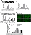
4p3f inhibits LPS-induced MMP-9 expression by reducing NF-κB activation. Cells were pretreated with Bay11-7082 (Bay, 0.1 μM) for 1 h and then incubated with LPS (10 μg/mL) for the indicated time intervals. (A) For MMP-9 expression, the conditioned media were collected and analyzed via gelatin zymography. The GAPDH level of cell lysates was assayed via Western blot. (B) For MMP-9 mRNA expression, total RNA was prepared and analyzed using real-time PCR. (C) Cells were pretreated with Bay (0.1 μM) or 4p3f (1 μM) for 1 h and then incubated with LPS (10 μg/mL) for the indicated times. After treatment, cell lysates were collected and analyzed using Western blot for phosphorylation of p65 NF-κB (p-p65) and GAPDH (as an internal control). (D) The translocation of p65 NF-κB was determined by means of immunofluorescence staining in RBAs. Cells were pretreated with Bay or 4p3f for 1 h, and then stimulated with LPS for 30 min. Cells were fixed and labeled with an anti-p65 NF-κB antibody and a FITC-conjugated secondary antibody. Individual cells were imaged (scale bar = 50 μm) as described in Section 2. (E) Cells were transiently transfected with a promoter–reporter construct (pGL4.32-containing κB binding sites), pretreated with Bay (0.1 μM) or 4p3f (1 μM) for 1 h, and then stimulated with LPS (10 μg/mL) for the indicated times. After stimulation, the firefly luciferase activity of the promoter (κB) construct was measured as relative promoter activity to that of Renilla luciferase activity. Data are expressed as mean ± SEM (n = 3). * p < 0.05 and ** p < 0.01, as compared with the untreated control; # p < 0.05 and ## p < 0.01, as compared with LPS-treated cells only. The images represent one of three individual experiments.
Figure 4
Figure 4
Effects of 4p3f on LPS-induced STAT3-mediated MMP-9 expression in RBAs. Cells were pretreated with or without STAT3I (1 μM) for 1 h and then incubated with LPS (10 μg/mL) for the indicated time intervals. (A) For MMP-9 expression, the conditioned media were collected and analyzed via gelatin zymography. The GAPDH level of cell lysates (as an internal control) was assayed by means of Western blot. (B) For MMP-9 mRNA expression, total RNA was prepared and analyzed via real-time PCR. (C) Cells were pretreated with STAT3I (1 μM) or 4p3f (1 μM) for 1 h and then incubated with LPS (10 μg/mL) for the indicated times. After treatment, cell lysates were collected and analyzed via Western blot for phosphorylation of STAT3 (p-STAT3) and total STAT3. Data are expressed as mean ± SEM (n = 3). * p < 0.05 and ** p < 0.01, as compared with the untreated control; # p < 0.05 and ## p < 0.01, as compared with LPS-treated cells only. The images represent one of three individual experiments.
Figure 5
Figure 5
MAPK-STAT3 signaling axis is crucial for LPS-stimulated NF-κB transcriptional activity in RBAs. Cells were pretreated with U0126 (0.1 μM), SB202190 (1 μM), or SP600125 (0.1 μM) for 1 h and then incubated with LPS (10 μg/mL) for the indicated times. After treatment, cell lysates were collected and analyzed via Western blot for phosphorylation of p65 NF-κB (p-p65) (A) and STAT3 (p-STAT3 and total STAT3) (B), as well as GAPDH (an internal control). (C) Cells were transiently transfected with a κB-Luc reporter construct (pGL4.32). After transfection, cells were pretreated with U0126 (0.1 μM), SB202190 (1 μM), SP600125 (0.1 μM), or STAT3I (1 μM) for 1 h and then incubated with LPS (10 μg/mL) for 4 h. Cell lysates were collected and detected using a Dual-Luciferase® Reporter Assay System. Data are expressed as mean ± SEM (n = 3). * p < 0.05 and ** p < 0.01, as compared with the untreated control; # p < 0.05 and ## p < 0.01, as compared with LPS-treated cells only. The images represent one of three individual experiments.
Figure 6
Figure 6
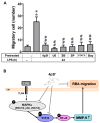
4p3f inhibits LPS-induced MMP-9-mediated astrocytic migration. (A) Cells were plated on 6-well culture plates, grew to confluence, and starved with a serum-free medium for 24 h. Cells were pretreated with U0126 (0.1 μM), SB202190 (1 μM), SP600125 (0.1 μM), STAT3I (1 μM), Bay (0.1 μM), or 4p3f (1 μM) for 1 h, and monolayer cells were manually scratched using a blue tip, as described in Section 2, and then incubated with LPS (10 μg/mL) for 24 h. Phase-contrast images of cells were taken at 24 h, and the number of cells that migrated was counted, as described in Section 2. Data are expressed as mean ± SEM (n = 3). ** p < 0.01, as compared with the untreated control; ## p < 0.01, as compared with LPS-treated cells only. (B) Schematic presentation of the effects of 4p3f on LPS-induced MMP-9 expression and astrocytic migration. In brain astrocytes (RBAs), LPS induces STAT3 activation through MAPK (i.e., ERK1/2, p38, and JNK1/2) signals, resulting in NF-κB-dependent MMP-9 expression. The increased MMP-9 expression leads to RBA migration. 4p3f inhibits these LPS-induced MMP-9-mediated events (brain astrocytic migration) by suppressing the activation of MAPKs, STAT3, and NF-κB signaling pathways.

Similar articles

References

    1. von Bartheld C.S., Bahney J., Herculano-Houzel S. The search for true numbers of neurons and glial cells in the human brain: A review of 150 years of cell counting. J. Comp. Neurol. 2016;524:3865–3895. doi: 10.1002/cne.24040. - DOI - PMC - PubMed
    1. Vivinetto A.L., Kim I.D., Goldberg D.C., Fones L., Brown E., Tarabykin V.S., Hill C.E., Cho S., Cave J.W. Zeb2 Is a Regulator of Astrogliosis and Functional Recovery after CNS Injury. Cell Rep. 2020;31:107834. doi: 10.1016/j.celrep.2020.107834. - DOI - PMC - PubMed
    1. Heithoff B.P., George K.K., Phares A.N., Zuidhoek I.A., Munoz-Ballester C., Robel S. Astrocytes are necessary for blood-brain barrier maintenance in the adult mouse brain. Glia. 2021;69:436–472. doi: 10.1002/glia.23908. - DOI - PMC - PubMed
    1. Faulkner J.R., Herrmann J.E., Woo M.J., Tansey K.E., Doan N.B., Sofroniew M.V. Reactive astrocytes protect tissue and preserve function after spinal cord injury. J. Neurosci. 2004;24:2143–2155. doi: 10.1523/JNEUROSCI.3547-03.2004. - DOI - PMC - PubMed
    1. Bush T.G., Puvanachandra N., Horner C.H., Polito A., Ostenfeld T., Svendsen C.N., Mucke L., Johnson M.H., Sofroniew M.V. Leukocyte infiltration, neuronal degeneration, and neurite outgrowth after ablation of scar-forming, reactive astrocytes in adult transgenic mice. Neuron. 1999;23:297–308. doi: 10.1016/S0896-6273(00)80781-3. - DOI - PubMed
  NODES
INTERN 7
twitter 2