Review
. 2024 Mar 30;8(1):19.
doi: 10.1038/s41538-024-00261-2.

Precision nutrition to reset virus-induced human metabolic reprogramming and dysregulation (HMRD) in long-COVID

Affiliations
Review

Precision nutrition to reset virus-induced human metabolic reprogramming and dysregulation (HMRD) in long-COVID

A Satyanarayan Naidu et al. NPJ Sci Food. .

Erratum in

Abstract

SARS-CoV-2, the etiological agent of COVID-19, is devoid of any metabolic capacity; therefore, it is critical for the viral pathogen to hijack host cellular metabolic machinery for its replication and propagation. This single-stranded RNA virus with a 29.9 kb genome encodes 14 open reading frames (ORFs) and initiates a plethora of virus-host protein-protein interactions in the human body. These extensive viral protein interactions with host-specific cellular _targets could trigger severe human metabolic reprogramming/dysregulation (HMRD), a rewiring of sugar-, amino acid-, lipid-, and nucleotide-metabolism(s), as well as altered or impaired bioenergetics, immune dysfunction, and redox imbalance in the body. In the infectious process, the viral pathogen hijacks two major human receptors, angiotensin-converting enzyme (ACE)-2 and/or neuropilin (NRP)-1, for initial adhesion to cell surface; then utilizes two major host proteases, TMPRSS2 and/or furin, to gain cellular entry; and finally employs an endosomal enzyme, cathepsin L (CTSL) for fusogenic release of its viral genome. The virus-induced HMRD results in 5 possible infectious outcomes: asymptomatic, mild, moderate, severe to fatal episodes; while the symptomatic acute COVID-19 condition could manifest into 3 clinical phases: (i) hypoxia and hypoxemia (Warburg effect), (ii) hyperferritinemia ('cytokine storm'), and (iii) thrombocytosis (coagulopathy). The mean incubation period for COVID-19 onset was estimated to be 5.1 days, and most cases develop symptoms after 14 days. The mean viral clearance times were 24, 30, and 39 days for acute, severe, and ICU-admitted COVID-19 patients, respectively. However, about 25-70% of virus-free COVID-19 survivors continue to sustain virus-induced HMRD and exhibit a wide range of symptoms that are persistent, exacerbated, or new 'onset' clinical incidents, collectively termed as post-acute sequelae of COVID-19 (PASC) or long COVID. PASC patients experience several debilitating clinical condition(s) with >200 different and overlapping symptoms that may last for weeks to months. Chronic PASC is a cumulative outcome of at least 10 different HMRD-related pathophysiological mechanisms involving both virus-derived virulence factors and a multitude of innate host responses. Based on HMRD and virus-free clinical impairments of different human organs/systems, PASC patients can be categorized into 4 different clusters or sub-phenotypes: sub-phenotype-1 (33.8%) with cardiac and renal manifestations; sub-phenotype-2 (32.8%) with respiratory, sleep and anxiety disorders; sub-phenotype-3 (23.4%) with skeleto-muscular and nervous disorders; and sub-phenotype-4 (10.1%) with digestive and pulmonary dysfunctions. This narrative review elucidates the effects of viral hijack on host cellular machinery during SARS-CoV-2 infection, ensuing detrimental effect(s) of virus-induced HMRD on human metabolism, consequential symptomatic clinical implications, and damage to multiple organ systems; as well as chronic pathophysiological sequelae in virus-free PASC patients. We have also provided a few evidence-based, human randomized controlled trial (RCT)-tested, precision nutrients to reset HMRD for health recovery of PASC patients.


Conflict of interest statement

The authors declare no competing interests.

Figures

Fig. 1
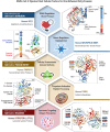
Fig. 1. Viral hijack of host cellular metabolic machinery.
SARS-CoV-2 infection of a susceptible host is achieved through viral spike (S)-protein-mediated hijack of human cell surface receptors (ACE2 and/or NRP1) and cell membrane proteases. The S1-region on viral S-protein contains a receptor-binding domain (RBD) that specifically recognizes host cell surface receptor(s) and exposes the S2 site. For fusion with host cell membrane, the viral S-protein hijacks specific cellular proteases for activation (‘priming’) of viral S-protein at the S1/S2 region. Subsequent conformational changes to viral S-protein lead to S1 shedding by cleavage of S1/S2 fragments. This process facilitates insertion of fusion peptide (FP) into host membrane. Accordingly, proteolytic cleavage by cellular enzymes TMPRSS2 and/or furin accomplish the task of viral FP insertion into host cell membrane. Alternatively, SARS-CoV-2 could also hijack lysosomal protease cathepsin L (CTSL) for direct viral endocytosis, where the viral membrane fuses with luminal face of the endosomal membrane facilitating viral RNA transfer into the cytosol. Thus, SARS-CoV-2 could infect the human by hijacking these 5 major host cellular factors via different routes of entry and elicit a wide range of clinical outcomes. The angiotensin-converting enzyme 2 (ACE2)/TMPRSS2-mediated viral infection and/or the ACE2/CTSL-mediated endosomal route may result in full-spectrum symptomatic COVID-19. The alternative neuropilin 1 (NRP1)/furin-mediated route, may down-regulate human pain receptors and manifest as asymptomatic to mild disease outcomes.
Fig. 2
Fig. 2. COVID-19 clinical spectrum.
The symptomatic outcomes of SARS-CoV-2 infection manifest in a tri-phasic manner as iron (Fe)-redox disruptive hematological syndromes. Phase-I: Hypoxia/Hypoxemia. Viral binding to ACE2 alters RAAS, subsequently lowers blood pressure, lung function, and reduces O2 transport (hypoxia) in the infected host. This condition triggers a mitochondrial metabolic shift by alteration of OXPHOS/TCA cycle and activation of anaerobic glycolysis, the ‘Warburg Effect’. This metabolic shift is regulated by HIF-1α that causes impairment of host immune response, exacerbates inflammation, and elicits tissue damage. This clinical phase of COVID-19 is considered a hypoxia-induced blood disease, associated with FeRD and HMRD,. Phase-II: Hyperferritinemia is characterized by a hyper-inflammatory state with elevated proinflammatory cytokines, which stimulates synthesis of both ferritin and hepcidin, the ultimate mediators of FeRD. The altered iron homeostasis is reflected by high iron content in reticuloendothelial cells and elevated serum ferritin levels. Such uncontrolled and dysfunctional immune response associated with macrophage activation leads to hyperferritinemia, and ‘cytokine storm’ or cytokine release syndrome (CRS). Hyperferritinemia, cellular redox imbalance and FeRD play a critical role in the disease progression of COVID-19,,. Phase-III: Thrombocytopenia. SARS-CoV-2 could invade blood vessels, induce vascular damage, and activate systemic thrombotic events with severe to fatal coagulopathies in COVID-19 patients. This clinical state along with hypoxia, could cast signs of hemolysis with release of heme proteins and accumulation of free heme. Heme from hemolysis could initiate oxidative and inflammatory stress that may cause microvascular thrombosis, organ ischemia and multi-organ failure in severe COVID-19 cases,.
Fig. 3
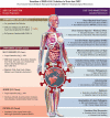
Fig. 3. Transition of SARS-CoV-2 Infection to Virus-free PASC: Pathophysiological Mechanisms.
Post-acute sequelae of COVID-19 (PASC) or long-COVID refers to a broad spectrum of symptoms and signs that are persistent, exacerbated, or new clinical incidents in the period that prolongs after acute SARS-CoV-2 infection. In acute COVID-19, the SARS‐CoV‐2 genome and its products critically reprogram and dysregulate human metabolism (HMRD) at transcription, translation, and post-translational modification (PTM) levels. Interaction of SARS-CoV-2 proteins with specific host cellular _targets rewires sugar-, amino acid-, lipid-, and nucleotide-metabolism(s), as well as alters or impairs bioenergetics, immune response, and redox homeostasis in the body, to facilitate viral replication and propagation,. However, several recoverees or survivors of COVID-19 (RT-PCR negative for SARS-CoV-2) continue to exhibit a plethora of clinical symptoms with impairment(s) of multiple organ systems. Accordingly, PASC or long-COVID is a virus-free, ‘new onset’ pathophysiological condition extending from a virus-induced HMRD. The HMRD in PASC pathology is a cumulative clinical outcome of several causative mechanisms comprising both SARS-CoV-2-derived virulence factors, as well as a multitude of host cellular factors and innate responses. A plethora of PASC clinical symptoms and related metabolic impairments indicate an involvement of different pathobiological mechanisms.
Fig. 4
Fig. 4. Virus-induced HMRD: hypoxia and ‘Warburg’ effect.
Dysregulation of glycolysis/TCA cycle is a key feature of HMRD. COVID-19 patients exhibit elevated serum glucose levels with an upregulation of glycolytic intermediates. Glutamine deficiency and hyaluronan over synthesis are HMRD-induced metabolic events in SARS-CoV-2 infection. M1 macrophages express nitric oxide synthase (NOS), which oxidizes arginine to nitric oxide (NO•) and citrulline. NO• modulates vascular tone, blood pressure and hemodynamics. Disrupted arginine metabolism further down-regulates NO• synthesis, aggravates endothelial dysfunction and triggers severe coagulopathies in COVID-19. Downstream generation of amino acids ornithine, citrulline, arginine in the circulation also indicates a severe renal dysfunction. Degradation of sphingomyelin by acid sphingomyelinase (ASM) generates stimulatory ceramides, the docking molecules for phospholipase A2 (PLA2). The hydrolysis of phospholipids (i.e., phosphatidyl choline) by PLA2 elevates arachidonic acid levels, a precursor for broad spectrum eicosanoids produced by cyclooxygenase (COX) and lipoxygenase (LOX) enzymes. These enzymes further convert arachidonic acid to prostaglandins (PGs), thromboxanes (TXs), and leukotrienes (LTs), which collectively contribute to the development of vascular inflammation and disease severity in COVID-19. Virus-induced HMRD alters host lipid metabolism with major impact on sphingolipid and arachidonic acid pathways. A decline in fat-soluble antioxidants’ vitamin E and carotenoids could compromises ROS quenching capacity in the plasma membrane, causes lipid peroxidation and OxS. Elevated serum lipase levels indicate damaging clinical outcomes in COVID-19 patients. The virus-induced HMRD alternations to glucose, amino acid, and lipid metabolism could aggravate the severity of COVID-19 and may extend to PASC pathology.
Fig. 5
Fig. 5. Viral-hijacked host cellular factors and consequences.
Clinical outcomes of COVID-19 depend on the ability of SARS-CoV-2 pathogen to hijack host metabolic machinery as well as cellular factors of an infected individual for invasion and internalization, followed by intra-cellular replication to assemble and release multiple viral copies for ultimate propagation/transmission. Each of the viral hijacked host cellular factor is also a quintessential functional component of human metabolism. Viral host receptor ACE2, is a critical regulator of blood pressure, controller of blood volume involved in systemic vascular resistance, and in CV homeostasis. Viral host receptor NRP1, is vital for several physiological pathways including nervous and vascular development, VEGF-dependent angiogenesis (i.e., new blood vessel formation), immunity and tumorigenesis,. Viral membrane fusion priming enzyme furin, is known for intracellular proteolytic processing of precursor polypeptides, which is an essential step in the maturation of many proteins such as plasma proteins, hormones, neuropeptides, and growth factors. Viral membrane fusion priming enzyme TMPRSS2 plays a key role in digestion, salt-water balance, iron metabolism, tissue remodeling, blood coagulation, auditory nerve development, and fertility. Viral endocytosis-mediator CTSL is involved in functional development of immune system, skeletal physiology including bone collagen degradation/resorption and thyroid hormone release,. Consequential to the viral hijack, these essential host cellular factors could malfunction and lead to a plethora of organ/system impairments with detrimental consequences to the human body. If not corrected or reset, this HMRD condition may persist in a PASC patient for weeks or months even after viral clearance and recovery from COVID-19.
Fig. 6
Fig. 6. Virus-induced HMRD of tryptophan metabolism/neuro-cognitive implications.
The SARS-CoV-2-induced HMR affects tryptophan metabolism by lowering the levels of tryptophan, serotonin, and indole-pyruvate, while elevating the levels of kynurenine, kynurenic acid, picolinic acid, and nicotinic acid,. After conversion to kynurenine, the tryptophan catabolism divides into different branches, leading to the formation of 3-OH-kynurenine, anthranilic acid or kynurenic acid. The 3-OH-kynurenine catabolism further leads to the generation of picolinic acid, quinolinic acid, and nicotinamide. The neuroprotective kynurenic acid is present mainly in astrocytes, neurotoxic 3-OH-kynurenine and excitotoxic quinolinic acid are found in microglial cells. Besides directly _targeting neurotransmitter receptors, the tryptophan metabolites, in particular 3-OH-kynurenine and 3-OH-anthranilic acid, are redox active that impact brain physiology. The modulation of the tryptophan-kynurenine pathway is an indicator for a coherent metabolic shift. The tryptophan-nicotinamide pathway is associated with inflammatory signals and coordinator of cell metabolism in SARS-CoV-2 infection. The broader virulence spectrum of SARS-CoV-2 with ability to cross the BBB and inflict a plethora of neuropathological manifestations by HMRD in host brain metabolism has been elucidated as ‘Neuro-COVID-19’.

Similar articles

Cited by

References

    1. Lu R, et al. Genomic characterisation and epidemiology of 2019 novel coronavirus: implications for virus origins and receptor binding. Lancet. 2020;395:565–574. doi: 10.1016/S0140-6736(20)30251-8. - DOI - PMC - PubMed
    1. Rajan, S. et al. In the wake of the pandemic: Preparing for long covid [internet]. National Center for Biotechnology Information Available at: https://pubmed.ncbi.nlm.nih.gov/33877759/ (Accessed: 27th March 2024).
    1. Coronavirus cases: Worldometer Available at: https://www.worldometers.info/coronavirus/ (Accessed: 27th March 2024).
    1. Hallek M, et al. Post-COVID syndrome. Dtsch. Arztebl. Int. 2023;120:48–55. - PMC - PubMed
    1. Davis HE, et al. Characterizing long COVID in an international cohort: 7 months of symptoms and their impact. EClinicalMedicine. 2021;38:101019. doi: 10.1016/j.eclinm.2021.101019. - DOI - PMC - PubMed
  NODES
Association 2
COMMUNITY 2
INTERN 11
twitter 2