Effects of Anti-Fibrotic Drugs on Transcriptome of Peripheral Blood Mononuclear Cells in Idiopathic Pulmonary Fibrosis
- PMID: 38612561
- PMCID: PMC11011476
- DOI: 10.3390/ijms25073750
Effects of Anti-Fibrotic Drugs on Transcriptome of Peripheral Blood Mononuclear Cells in Idiopathic Pulmonary Fibrosis
Abstract
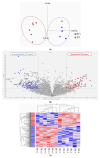
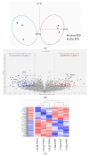
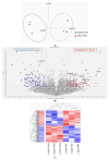
Two anti-fibrotic drugs, pirfenidone (PFD) and nintedanib (NTD), are currently used to treat idiopathic pulmonary fibrosis (IPF). Peripheral blood mononuclear cells (PBMCs) are immunocompetent cells that could orchestrate cell-cell interactions associated with IPF pathogenesis. We employed RNA sequencing to examine the transcriptome signature in the bulk PBMCs of patients with IPF and the effects of anti-fibrotic drugs on these signatures. Differentially expressed genes (DEGs) between "patients with IPF and healthy controls" and "before and after anti-fibrotic treatment" were analyzed. Enrichment analysis suggested that fatty acid elongation interferes with TGF-β/Smad signaling and the production of oxidative stress since treatment with NTD upregulates the fatty acid elongation enzymes ELOVL6. Treatment with PFD downregulates COL1A1, which produces wound-healing collagens because activated monocyte-derived macrophages participate in the production of collagen, type I, and alpha 1 during tissue damage. Plasminogen activator inhibitor-1 (PAI-1) regulates wound healing by inhibiting plasmin-mediated matrix metalloproteinase activation, and the inhibition of PAI-1 activity attenuates lung fibrosis. DEG analysis suggested that both the PFD and NTD upregulate SERPINE1, which regulates PAI-1 activity. This study embraces a novel approach by using RNA sequencing to examine PBMCs in IPF, potentially revealing systemic biomarkers or pathways that could be _targeted for therapy.
Keywords: RNA sequencing; idiopathic pulmonary fibrosis; nintedanib; peripheral blood mononuclear cells (PBMCs); pirfenidone; transcriptome.
Conflict of interest statement
Mitsuhiro Abe reports personal speaking and lecture fees from Boehringer Ingelheim, Japan. Takuji Suzuki reports on the personal speaking and lecture fees received from Boehringer Ingelheim, Japan.
Figures


Similar articles
-
Design, synthesis, and evaluation of pirfenidone-NSAIDs conjugates for the treatment of idiopathic pulmonary fibrosis.Bioorg Chem. 2024 Feb;143:107018. doi: 10.1016/j.bioorg.2023.107018. Epub 2023 Dec 6. Bioorg Chem. 2024. PMID: 38071874
-
Efficacy of antifibrotic drugs, nintedanib and pirfenidone, in treatment of progressive pulmonary fibrosis in both idiopathic pulmonary fibrosis (IPF) and non-IPF: a systematic review and meta-analysis.BMC Pulm Med. 2021 Dec 11;21(1):411. doi: 10.1186/s12890-021-01783-1. BMC Pulm Med. 2021. PMID: 34895203 Free PMC article.
-
Plasminogen activator inhibitor-1 suppresses profibrotic responses in fibroblasts from fibrotic lungs.J Biol Chem. 2015 Apr 10;290(15):9428-41. doi: 10.1074/jbc.M114.601815. Epub 2015 Feb 3. J Biol Chem. 2015. PMID: 25648892 Free PMC article.
-
Nintedanib for the treatment of idiopathic pulmonary fibrosis.Expert Opin Pharmacother. 2018 Feb;19(2):167-175. doi: 10.1080/14656566.2018.1425681. Epub 2018 Jan 12. Expert Opin Pharmacother. 2018. PMID: 29327616 Review.
-
Newer developments in idiopathic pulmonary fibrosis in the era of anti-fibrotic medications.Expert Rev Respir Med. 2016 Jun;10(6):699-711. doi: 10.1080/17476348.2016.1177461. Epub 2016 Apr 26. Expert Rev Respir Med. 2016. PMID: 27094006 Review.
References
-
- Raghu G., Remy-Jardin M., Myers J.L., Richeldi L., Ryerson C.J., Lederer D.J., Behr J., Cottin V., Danoff S.K., Morell F., et al. Diagnosis of Idiopathic Pulmonary Fibrosis. An Official ATS/ERS/JRS/ALAT Clinical Practice Guideline. Am. J. Respir. Crit. Care Med. 2018;198:e44–e68. doi: 10.1164/rccm.201807-1255ST. - DOI - PubMed
-
- Oku H., Shimizu T., Kawabata T., Nagira M., Hikita I., Ueyama A., Matsushima S., Torii M., Arimura A. Antifibrotic action of pirfenidone and prednisolone: Different effects on pulmonary cytokines and growth factors in bleomycin-induced murine pulmonary fibrosis. Eur. J. Pharmacol. 2008;590:400–408. doi: 10.1016/j.ejphar.2008.06.046. - DOI - PubMed
MeSH terms
Substances
Grants and funding
LinkOut - more resources
Full Text Sources
Miscellaneous

