Exploring morphological similarity and randomness in Alzheimer's disease using adjacent grey matter voxel-based structural analysis
- PMID: 38654366
- PMCID: PMC11036786
- DOI: 10.1186/s13195-024-01448-1
Exploring morphological similarity and randomness in Alzheimer's disease using adjacent grey matter voxel-based structural analysis
Abstract
Background: Alzheimer's disease is characterized by large-scale structural changes in a specific pattern. Recent studies developed morphological similarity networks constructed by brain regions similar in structural features to represent brain structural organization. However, few studies have used local morphological properties to explore inter-regional structural similarity in Alzheimer's disease.
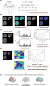
Methods: Here, we sourced T1-weighted MRI images of 342 cognitively normal participants and 276 individuals with Alzheimer's disease from the Alzheimer's Disease Neuroimaging Initiative database. The relationships of grey matter intensity between adjacent voxels were defined and converted to the structural pattern indices. We conducted the information-based similarity method to evaluate the structural similarity of structural pattern organization between brain regions. Besides, we examined the structural randomness on brain regions. Finally, the relationship between the structural randomness and cognitive performance of individuals with Alzheimer's disease was assessed by stepwise regression.
Results: Compared to cognitively normal participants, individuals with Alzheimer's disease showed significant structural pattern changes in the bilateral posterior cingulate gyrus, hippocampus, and olfactory cortex. Additionally, individuals with Alzheimer's disease showed that the bilateral insula had decreased inter-regional structural similarity with frontal regions, while the bilateral hippocampus had increased inter-regional structural similarity with temporal and subcortical regions. For the structural randomness, we found significant decreases in the temporal and subcortical areas and significant increases in the occipital and frontal regions. The regression analysis showed that the structural randomness of five brain regions was correlated with the Mini-Mental State Examination scores of individuals with Alzheimer's disease.
Conclusions: Our study suggested that individuals with Alzheimer's disease alter micro-structural patterns and morphological similarity with the insula and hippocampus. Structural randomness of individuals with Alzheimer's disease changed in temporal, frontal, and occipital brain regions. Morphological similarity and randomness provide valuable insight into brain structural organization in Alzheimer's disease.
Keywords: Alzheimer’s disease; Information-based similarity method; Morphological similarity network; Structural magnetic resonance imaging.
© 2024. The Author(s).
Conflict of interest statement
The authors declare no competing interests.
Figures


Similar articles
-
The Neuropsychological Correlates of Brain Perfusion and Gray Matter Volume in Alzheimer's Disease.J Alzheimers Dis. 2020;78(4):1639-1652. doi: 10.3233/JAD-200676. J Alzheimers Dis. 2020. PMID: 33185599
-
Morphological and Structural Network Analysis of Sporadic Alzheimer's Disease Brains Based on the APOE4 Gene.J Alzheimers Dis. 2023;91(3):1035-1048. doi: 10.3233/JAD-220877. J Alzheimers Dis. 2023. PMID: 36530087
-
Structural neuroimaging changes associated with subjective cognitive decline from a clinical sample.Neuroimage Clin. 2024;42:103615. doi: 10.1016/j.nicl.2024.103615. Epub 2024 May 10. Neuroimage Clin. 2024. PMID: 38749146 Free PMC article.
-
Detection of grey matter loss in mild Alzheimer's disease with voxel based morphometry.J Neurol Neurosurg Psychiatry. 2002 Dec;73(6):657-64. doi: 10.1136/jnnp.73.6.657. J Neurol Neurosurg Psychiatry. 2002. PMID: 12438466 Free PMC article.
-
[Neuroimaging in Alzheimer's disease: a synthesis and a contribution to the understanding of physiopathological mechanisms].Biol Aujourdhui. 2010;204(2):145-58. doi: 10.1051/jbio/2010010. Epub 2010 Jun 21. Biol Aujourdhui. 2010. PMID: 20950559 Review. French.
References
-
- Du AT, Schuff N, Amend D, Laakso MP, Hsu YY, Jagust WJ, Yaffe K, Kramer JH, Reed B, Norman D, et al. Magnetic resonance imaging of the entorhinal cortex and hippocampus in mild cognitive impairment and Alzheimer's disease. J Neurol Neurosurg Psychiatry. 2001;71(4):441–447. doi: 10.1136/jnnp.71.4.441. - DOI - PMC - PubMed
-
- Buckner RL, Snyder AZ, Shannon BJ, LaRossa G, Sachs R, Fotenos AF, Sheline YI, Klunk WE, Mathis CA, Morris JC, et al. Molecular, Structural, and Functional Characterization of Alzheimer's Disease: Evidence for a Relationship between Default Activity, Amyloid, and Memory. J Neurosci. 2005;25(34):7709–7717. doi: 10.1523/JNEUROSCI.2177-05.2005. - DOI - PMC - PubMed
Publication types
MeSH terms
Grants and funding
- MOHW112-CDC-C-114-000111/Taiwan Centers for Disease Control
- Mt. Jade Young Scholarship Award/Ministry of Education
- 112W32101/Brain Research Center, National Yang Ming Chiao Tung University
- 112-2823-8-A49-001, 111-2634-F-A49-014, 112-2321-B-A49-013, 112-2321-B-A49-021, and 112-2634-F-A49-003/National Science and Technology Council
- V112C-054, V113C-144, and V113E-008-3/Taipei Veterans General Hospital
LinkOut - more resources
Full Text Sources
Medical

