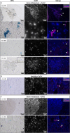
Magnetic Particle Imaging Reveals that Iron-Labeled Extracellular Vesicles Accumulate in Brains of Mice with Metastases
- PMID: 38860682
- PMCID: PMC11194773
- DOI: 10.1021/acsami.4c04920
Magnetic Particle Imaging Reveals that Iron-Labeled Extracellular Vesicles Accumulate in Brains of Mice with Metastases
Abstract
The incidence of breast cancer remains high worldwide and is associated with a significant risk of metastasis to the brain that can be fatal; this is due, in part, to the inability of therapeutics to cross the blood-brain barrier (BBB). Extracellular vesicles (EVs) have been found to cross the BBB and further have been used to deliver drugs to tumors. EVs from different cell types appear to have different patterns of accumulation and retention as well as the efficiency of bioactive cargo delivery to recipient cells in the body. Engineering EVs as delivery tools to treat brain metastases, therefore, will require an understanding of the timing of EV accumulation and their localization relative to metastatic sites. Magnetic particle imaging (MPI) is a sensitive and quantitative imaging method that directly detects superparamagnetic iron. Here, we demonstrate MPI as a novel tool to characterize EV biodistribution in metastatic disease after labeling EVs with superparamagnetic iron oxide (SPIO) nanoparticles. Iron-labeled EVs (FeEVs) were collected from iron-labeled parental primary 4T1 tumor cells and brain-seeking 4T1BR5 cells, followed by injection into the mice with orthotopic tumors or brain metastases. MPI quantification revealed that FeEVs were retained for longer in orthotopic mammary carcinomas compared to SPIOs. MPI signal due to iron could only be detected in brains of mice bearing brain metastases after injection of FeEVs, but not SPIOs, or FeEVs when mice did not have brain metastases. These findings indicate the potential use of EVs as a therapeutic delivery tool in primary and metastatic tumors.
Keywords: brain metastasis; breast cancer; exosome; extracellular vesicles; magnetic particle imaging.
Conflict of interest statement
The authors declare no competing financial interest.
Figures


Similar articles
-
Magnetic Particle Imaging of Macrophages Associated with Cancer: Filling the Voids Left by Iron-Based Magnetic Resonance Imaging.Mol Imaging Biol. 2020 Aug;22(4):958-968. doi: 10.1007/s11307-020-01473-0. Mol Imaging Biol. 2020. PMID: 31933022
-
Comparison between USPIOs and SPIOs for Multimodal Imaging of Extracellular Vesicles Extracted from Adipose Tissue-Derived Adult Stem Cells.Int J Mol Sci. 2024 Sep 7;25(17):9701. doi: 10.3390/ijms25179701. Int J Mol Sci. 2024. PMID: 39273647 Free PMC article.
-
Sensitive and specific detection of breast cancer lymph node metastasis through dual-modality magnetic particle imaging and fluorescence molecular imaging: a preclinical evaluation.Eur J Nucl Med Mol Imaging. 2022 Jul;49(8):2723-2734. doi: 10.1007/s00259-022-05834-5. Epub 2022 May 20. Eur J Nucl Med Mol Imaging. 2022. PMID: 35590110 Free PMC article.
-
Magnetic iron oxide nanoparticles for imaging, _targeting and treatment of primary and metastatic tumors of the brain.J Control Release. 2020 Apr 10;320:45-62. doi: 10.1016/j.jconrel.2020.01.009. Epub 2020 Jan 7. J Control Release. 2020. PMID: 31923537 Free PMC article. Review.
-
_targeted extracellular vesicle delivery systems employing superparamagnetic iron oxide nanoparticles.Acta Biomater. 2021 Oct 15;134:13-31. doi: 10.1016/j.actbio.2021.07.027. Epub 2021 Jul 18. Acta Biomater. 2021. PMID: 34284151 Review.
Cited by
-
Surface Engineering of Magnetic Iron Oxide Nanoparticles for Breast Cancer Diagnostics and Drug Delivery.Int J Nanomedicine. 2024 Aug 17;19:8437-8461. doi: 10.2147/IJN.S477652. eCollection 2024. Int J Nanomedicine. 2024. PMID: 39170101 Free PMC article. Review.
References
-
- Ferlay J.; Ervik M.; Laversanne M.; Colombet M.; Mery L.; Piñeros M.; Znaor A.; Soerjomataram I.; Bray F.. Global Cancer Observatory: Cancer Today; International Agency for Research on Cancer. https://gco.iarc.who.int/today (accessed May 22, 2024).
MeSH terms
Substances
LinkOut - more resources
Full Text Sources
Medical

