. 2024 Jul 1:39:101768.
doi: 10.1016/j.bbrep.2024.101768. eCollection 2024 Sep.

Proteolysis of mitochondrial calpain-13 in cerebral ischemia-reperfusion injury

Affiliations

Proteolysis of mitochondrial calpain-13 in cerebral ischemia-reperfusion injury

Yusaku Chukai et al. Biochem Biophys Rep. .

Abstract

Calpains are calcium-dependent cysteine proteases activated by intracellular Ca2+. Although calpains mainly exist in the cytosol, calpain-13 is present in the mitochondria in mouse brains; however, the enzymatic properties and physiological functions of calpain-13 remain unknown. Hence, in this study, we predicted and evaluated the enzymatic properties of calpain-13. Based on our bioinformatic approaches, calpain-13 possessed a catalytic triad and EF-hand domain, similar to calpain-1, a well-studied calpain. Therefore, we hypothesized that calpain-13 had calpain-1-like enzymatic properties; however, calpain-13 was not proteolyzed in C57BL/6J mouse brains. Subsequently, cerebral ischemia/reperfusion (I/R) injury caused proteolysis of mitochondrial calpain-13. Thus, our study showed that mitochondrial calpain-13 was proteolyzed in the mitochondria of the I/R injured mouse brain. This finding could be valuable in further research elucidating the involvement of calpain-13 in cell survival or death in brain diseases, such as cerebral infarction.

Keywords: Calcium; Calpain catalytic domain; Calpain-13; Cerebral ischemia/reperfusion injury; Mitochondrion; Mouse brain.


Conflict of interest statement

The authors declare that they have no known competing financial interests or personal relationships that could have appeared to influence the work reported in this paper.

Figures

Image 1
Graphical abstract
Fig. 1
Fig. 1
Comparison of amino acid sequences between mouse calpain-1 (CAPN1) and -13 (CAPN13). The domains of CAPN1 are shown under the amino acid sequences. White characters in the red box indicate the completely conserved residues, whereas red characters in the blue box indicate similar residues. White arrowheads indicate the three conserved residues in the active site. (For interpretation of the references to color in this figure legend, the reader is referred to the Web version of this article.)
Fig. 2
Fig. 2
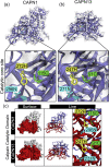
Structural analysis of the catalytic core site and EF-hand domains of mouse calpains-1 and -13. (a and b) The predicted structure of mouse calpain-1 (CAPN1) and -13 (CAPN13). The catalytic triad is preserved in both calpains. (c) The calpain catalytic core site of calpains-1 and -13 is in the inner region of the structure. The red color indicates the calpain catalytic domain. Green, cysteine (C); yellow, histidine (H); blue, asparagine (N). (d and e) The EF-hand domain is in a similar position in calpain-1 and -13. The red color indicates the EF-hand domain 2 from 534F to 569C (d) and domain 4 from 632I to 665S (e) in calpain-13. (For interpretation of the references to color in this figure legend, the reader is referred to the Web version of this article.)
Fig. 2
Fig. 2
Structural analysis of the catalytic core site and EF-hand domains of mouse calpains-1 and -13. (a and b) The predicted structure of mouse calpain-1 (CAPN1) and -13 (CAPN13). The catalytic triad is preserved in both calpains. (c) The calpain catalytic core site of calpains-1 and -13 is in the inner region of the structure. The red color indicates the calpain catalytic domain. Green, cysteine (C); yellow, histidine (H); blue, asparagine (N). (d and e) The EF-hand domain is in a similar position in calpain-1 and -13. The red color indicates the EF-hand domain 2 from 534F to 569C (d) and domain 4 from 632I to 665S (e) in calpain-13. (For interpretation of the references to color in this figure legend, the reader is referred to the Web version of this article.)
Fig. 3
Fig. 3
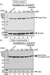
In vitro calcium assay of cytosolic and mitochondrial calpain-13 in the mouse brain. (a and b) In vitro assay for Ca2+ concentration-dependency for calpain-13 in cytosol (a) and mitochondria (b). Each sample (cytosol, 20 μg/lane; mitochondria, 40 μg/lane) was incubated with 0–1000 μM Ca2+ concentrations for 60 min at 37 °C. (c and d) In vitro assay for time-dependency for calpain-13 in cytosol (c) and mitochondria (d). Each sample (cytosol, 20 μg/lane; mitochondria, 40 μg/lane) was incubated with 1 mM Ca2+ for 0–60 min at 37 °C. β-Actin and Adenylate kinase 2 (AK2) were used as the cytosolic and mitochondrial loading control. M, molecular weight marker. “No incu.” indicates the untreated sample.
Fig. 3
Fig. 3
In vitro calcium assay of cytosolic and mitochondrial calpain-13 in the mouse brain. (a and b) In vitro assay for Ca2+ concentration-dependency for calpain-13 in cytosol (a) and mitochondria (b). Each sample (cytosol, 20 μg/lane; mitochondria, 40 μg/lane) was incubated with 0–1000 μM Ca2+ concentrations for 60 min at 37 °C. (c and d) In vitro assay for time-dependency for calpain-13 in cytosol (c) and mitochondria (d). Each sample (cytosol, 20 μg/lane; mitochondria, 40 μg/lane) was incubated with 1 mM Ca2+ for 0–60 min at 37 °C. β-Actin and Adenylate kinase 2 (AK2) were used as the cytosolic and mitochondrial loading control. M, molecular weight marker. “No incu.” indicates the untreated sample.
Fig. 4
Fig. 4
In vivo proteolysis assay of cytosolic and mitochondrial calpain-13 using a cerebral I/R injury mouse model. (a) Western blot analysis of the proteolysis/autolysis products of cytosolic calpain-13 after 15 min of ischemia and 3–24 h of reperfusion (20 μg/lane). β-Actin was used as a cytosolic loading control. (b) Western blot analysis of the proteolysis/autolysis products of mitochondrial calpain-13 after 15 min of ischemia and 3–24 h of reperfusion (40 μg/lane). Cytochrome c oxidase subunit 4 (COX IV) was used as a mitochondrial loading control. M, molecular weight marker. (c) Statistical analysis of the proteolytic form of mitochondrial calpain-13 after 15 min of ischemia and 24 h of reperfusion (I/R). The expression is significantly increased in I/R mice compared to that in the sham control (*p = 0.041). Data indicate the average ± standard deviation of each group, and each dot represents the individual value. Detailed statistics are provided in Table S1.

Similar articles

References

    1. Goll D.E., Thompson V.F., Li H., Wei W., Cong J. The calpain system. Physiol. Rev. 2003;83:731–801. doi: 10.1152/PHYSREV.00029.2002. - DOI - PubMed
    1. Smith M.A., Schnellmann R.G. Calpains, mitochondria, and apoptosis. Cardiovasc. Res. 2012;96:32–37. doi: 10.1093/CVR/CVS163. - DOI - PMC - PubMed
    1. Hosseini M., Najmabadi H., Kahrizi K. Calpains: diverse functions but enigmatic, arch. Iran. Méd. 2018;21:170–179. - PubMed
    1. Ono Y., Sorimachi H. Calpains: an elaborate proteolytic system. Biochim. Biophys. Acta. 2012;1824:224–236. doi: 10.1016/J.BBAPAP.2011.08.005. - DOI - PubMed
    1. Ozaki T., Tomita H., Tamai M., Ishiguro S.I. Characteristics of mitochondrial calpains. J. Biochem. 2007;142:365–376. doi: 10.1093/JB/MVM143. - DOI - PubMed

LinkOut - more resources

  NODES
twitter 2