Review
. 2024 Oct 7;29(19):4737.
doi: 10.3390/molecules29194737.

Recent Advances and Prospects of Nucleic Acid Therapeutics for Anti-Cancer Therapy

Affiliations
Review

Recent Advances and Prospects of Nucleic Acid Therapeutics for Anti-Cancer Therapy

Minhyuk Lee et al. Molecules. .

Abstract

Nucleic acid therapeutics are promising alternatives to conventional anti-cancer therapy, such as chemotherapy and radiation therapy. While conventional therapies have limitations, such as high side effects, low specificity, and drug resistance, nucleic acid therapeutics work at the gene level to eliminate the cause of the disease. Nucleic acid therapeutics treat diseases in various forms and using different mechanisms, including plasmid DNA (pDNA), small interfering RNA (siRNA), anti-microRNA (anti-miR), microRNA mimics (miRNA mimic), messenger RNA (mRNA), aptamer, catalytic nucleic acid (CNA), and CRISPR cas9 guide RNA (gRNA). In addition, nucleic acids have many advantages as nanomaterials, such as high biocompatibility, design flexibility, low immunogenicity, small size, relatively low price, and easy functionalization. Nucleic acid therapeutics can have a high therapeutic effect by being used in combination with various nucleic acid nanostructures, inorganic nanoparticles, lipid nanoparticles (LNPs), etc. to overcome low physiological stability and cell internalization efficiency. The field of nucleic acid therapeutics has advanced remarkably in recent decades, and as more and more nucleic acid therapeutics have been approved, they have already demonstrated their potential to treat diseases, including cancer. This review paper introduces the current status and recent advances in nucleic acid therapy for anti-cancer treatment and discusses the tasks and prospects ahead.

Keywords: RNA interference (RNAi); anti-cancer therapy; aptamer; gene therapy; nucleic acid delivery; nucleic acid therapeutics (NATs).


Conflict of interest statement

The authors declare no conflicts of interest.

Figures

Figure 5
Figure 5
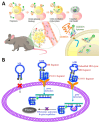
Anti-cancer therapeutic strategies using aptamer-based protein inhibition. (A) Schematic image of multimeric aptamer for EpCAM and PDGF protein inhibition. (B) Schematic image of bispecific aptamer for lysosomal degradation of _target membrane proteins with IGFIIR-mediated lysosomal proteolysis systems. (C) Schematic image of bispecific aptamer for _target membrane receptor inhibition by thrombin-mediated cleavage. (Adapted from Refs. [134,138,139]).
Figure 6
Figure 6
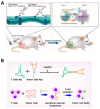
Anti-cancer therapeutic strategies using aptamer-based immunotherapy. (A) Schematic image of aptamer-based immunotherapy that induces apoptosis of cancer cells by blocking immune checkpoints. (B) Schematic image of bispecific aptamer that mediates cancer cell recognition of T cells. (Adapted from Refs. [146,149]).
Figure 7
Figure 7
Anti-cancer therapeutic strategies using aptamer-based _targeted therapy. (A) Schematic image of 5-Fu conjugated CD44E/s bispecific aptamer-based therapeutic for _targeted delivery of anti-cancer drugs. (B) Schematic image of DNAzyme-embedded AS1411 aptamer that simultaneously detects and suppresses tumor genes. (Adapted from Refs. [154,163]).
Figure 1
Figure 1
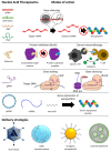
Overview of types, modes of action, and delivery strategies of nucleic acid-based therapeutics for anti-cancer therapy. siRNA, small interfering RNA; miR, microRNA; ASO, antisense oligonucleotide; CNA, catalytic nucleic acid; gRNA, guide RNA; pDNA, plasmid DNA; mRNA, messenger RNA.
Figure 2
Figure 2
(A) Schematic image of gene silencing with siRNA or miR mimic. The siRNA and miR mimic are incorporated within the RISC in the cytoplasm and then unwind, activating the guide strands. (B) Schematic image of gene silencing with RNase H-mediated cleavage with ASO. ASOs with chemically modified sugar backbones inhibit mRNA expression through steric hindrance. (C) Schematic image of gene silencing with CNA-based cleavage. The presence of metal ions helps to stabilize the activated structure of ribozymes and is essential for the catalytic activity of DNAzymes.
Figure 3
Figure 3
Schematic image of the mechanism of anti-miRs inducing upregulation of tumor suppressor genes by blocking oncogenic miRISC. Oncogenic miRICS inhibits expression by cleaving tumor suppressor-encoded mRNAs. Anti-miRs hybridize to oncogenic miRICS and block the gene silencing activity. As a result, tumor suppressor genes are upregulated.
Figure 4
Figure 4
Schematic images of the secondary structure of (A) the hammerhead ribozyme–substrate complex and (B) the 10–23 DNAzyme–substrate complex. The red arrow indicates the cleavage site of the substrate RNA. (A) The hammerhead ribozyme consists of an intrastrand helix (helix II) and two interstrand helixes (helix I and III) generated after hybridizing with substrate RNA and a conserved unpaired catalytic core. (B) The 10–23 DNAzyme consists of two variable binding arms, I and II, on both sides of the conserved (15 nt) and unpaired catalytic core. R represents A or G; Y represents U (or T) or C; H represents A, C, or U; N represents A, U (or T), C, or G.
Figure 8
Figure 8
Schematic images of the CRISPR Cas9 and CRISPR dCas9 systems. (A) Schematic image of CRISPR Cas9 genome editing mechanism. The endonuclease domains of Cas9 (RuvC and HNH) cleave the _target sequence after hybridization, and the cut DNA is repaired by NHEJ or HDR, which results in gene disruption or replacement. (B) Schematic image of CRISPR dCas9 gene-silencing mechanism. Mutations in the nuclease domain of dCas9 (D10A and H840A) render it unable to cleave the _target sequence after hybridization and interfere with the RNA transcription process through steric hindrance.
Figure 9
Figure 9
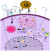
Schematic image of the anti-cancer mechanisms of pDNA and mRNA therapeutics. pDNA is delivered to the nucleus and replicated or transcribed by enzymes to synthesize RNA with therapeutic effects, such as gRNA, pre-mRNA, or shRNA, depending on the genes inserted. Pre-mRNA and shRNA are post-transcriptionally processed by enzymes into mRNA and siRNA, respectively, and transported out of the nucleus. mRNA is transported into the cytoplasm and translated by ribosomes to synthesize the encoded proteins, such as antigens, antibodies, cytokines, tumor suppressor proteins, and Cas9 proteins. The numbers represent (1) expression of a protein with anti-cancer effects, (2) DNA vaccine that induces immunotherapy by expressing an antigen or checkpoint inhibitor, and (3) expression of RNA-based NATs.

Similar articles

References

    1. Gopal S., Sivaram S., Rajaraman P., Trimble E.L. Think globally about cancer. Nat. Med. 2019;25:351. - PubMed
    1. Ferlay J., Colombet M., Soerjomataram I., Parkin D.M., Piñeros M., Znaor A., Bray F. Cancer statistics for the year 2020: An overview. Int. J. Cancer. 2021;149:778–789. doi: 10.1002/ijc.33588. - DOI - PubMed
    1. Zhang L., Lou W., Wang J. Advances in nucleic acid therapeutics: Structures, delivery systems, and future perspectives in cancer treatment. Clin. Exp. Med. 2024;24:200. doi: 10.1007/s10238-024-01463-4. - DOI - PMC - PubMed
    1. Yahya E.B., Alqadhi A.M. Recent trends in cancer therapy: A review on the current state of gene delivery. Life Sci. 2021;269:119087. doi: 10.1016/j.lfs.2021.119087. - DOI - PubMed
    1. Das S.K., Menezes M.E., Bhatia S., Wang X., Emdad L., Sarkar D., Fisher P.B. Gene Therapies for Cancer: Strategies, Challenges and Successes. J. Cell. Physiol. 2014;230:259–271. doi: 10.1002/jcp.24791. - DOI - PMC - PubMed

LinkOut - more resources

  NODES
HOME 2
Intern 1
Javascript 1
mac 3
os 19
text 12
twitter 2
web 4