Mouse Zic1 is involved in cerebellar development
- PMID: 9412507
- PMCID: PMC6793425
- DOI: 10.1523/JNEUROSCI.18-01-00284.1998
Mouse Zic1 is involved in cerebellar development
Abstract
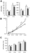
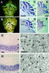
Zic genes encode zinc finger proteins, the expression of which is highly restricted to cerebellar granule cells and their precursors. These genes are homologs of the Drosophila pair-rule gene odd-paired. To clarify the role of the Zic1 gene, we have generated mice deficient in Zic1. Homozygous mice showed remarkable ataxia during postnatal development. Nearly all of the mice died within 1 month. Their cerebella were hypoplastic and missing a lobule in the anterior lobe. A bromodeoxyuridine labeling study indicated a reduction both in the proliferating cell fraction in the external germinal layer (EGL), from 14 d postcoitum, and in forward movement of the EGL. These findings suggest that Zic1 may determine the cerebellar folial pattern principally via regulation of cell proliferation in the EGL.
Figures


Similar articles
-
Zic2 controls cerebellar development in cooperation with Zic1.J Neurosci. 2002 Jan 1;22(1):218-25. doi: 10.1523/JNEUROSCI.22-01-00218.2002. J Neurosci. 2002. PMID: 11756505 Free PMC article.
-
Identification and characterization of Zic4, a new member of the mouse Zic gene family.Gene. 1996 Jun 26;172(2):291-4. doi: 10.1016/0378-1119(96)00111-4. Gene. 1996. PMID: 8682319
-
The expression of the mouse Zic1, Zic2, and Zic3 gene suggests an essential role for Zic genes in body pattern formation.Dev Biol. 1997 Feb 15;182(2):299-313. doi: 10.1006/dbio.1996.8449. Dev Biol. 1997. PMID: 9070329
-
ZIC1 Function in Normal Cerebellar Development and Human Developmental Pathology.Adv Exp Med Biol. 2018;1046:249-268. doi: 10.1007/978-981-10-7311-3_13. Adv Exp Med Biol. 2018. PMID: 29442326 Review.
-
Odd-Paired: The Drosophila Zic Gene.Adv Exp Med Biol. 2018;1046:41-58. doi: 10.1007/978-981-10-7311-3_3. Adv Exp Med Biol. 2018. PMID: 29442316 Review.
Cited by
-
Absence of an external germinal layer in zebrafish and shark reveals a distinct, anamniote ground plan of cerebellum development.J Neurosci. 2010 Feb 24;30(8):3048-57. doi: 10.1523/JNEUROSCI.6201-09.2010. J Neurosci. 2010. PMID: 20181601 Free PMC article.
-
Prenatal Exposure to Paint Thinner Alters Postnatal Development and Behavior in Mice.Front Behav Neurosci. 2017 Sep 11;11:171. doi: 10.3389/fnbeh.2017.00171. eCollection 2017. Front Behav Neurosci. 2017. PMID: 28959195 Free PMC article.
-
The ZIC gene family encodes multi-functional proteins essential for patterning and morphogenesis.Cell Mol Life Sci. 2013 Oct;70(20):3791-811. doi: 10.1007/s00018-013-1285-5. Epub 2013 Feb 27. Cell Mol Life Sci. 2013. PMID: 23443491 Free PMC article. Review.
-
Dual control of neurogenesis by PC3 through cell cycle inhibition and induction of Math1.J Neurosci. 2004 Mar 31;24(13):3355-69. doi: 10.1523/JNEUROSCI.3860-03.2004. J Neurosci. 2004. PMID: 15056715 Free PMC article.
-
Combined overexpression of ATXN1L and mutant ATXN1 knockdown by AAV rescue motor phenotypes and gene signatures in SCA1 mice.Mol Ther Methods Clin Dev. 2022 Apr 12;25:333-343. doi: 10.1016/j.omtm.2022.04.004. eCollection 2022 Jun 9. Mol Ther Methods Clin Dev. 2022. PMID: 35573049 Free PMC article.
References
-
- Alder J, Cho NK, Hatten ME. Embryonic precursor cells from the rhombic lip are specified to a cerebellar granule neuron identity. Neuron. 1996;17:389–399. - PubMed
-
- Altman J. Postnatal development of the cerebellar cortex in the rat. II. Phases in the maturation of Purkinje cells and of the molecular layer. J Comp Neurol. 1972;145:399–463. - PubMed
-
- Altman J, Anderson WJ. Experimental reorganization of the cerebellar cortex: I. Morphological effects of elimination of all micro neurons with prolonged x-irradiation started at birth. J Comp Neurol. 1972;146:355–406. - PubMed
-
- Altman J, Bayer SA. Development of the cerebellar system. CRC; New York: 1996.
-
- Aruga J, Yokota N, Hashimoto M, Furuichi T, Fukuda M, Mikoshiba K. A novel zinc finger protein, Zic, is involved in neurogenesis, especially in the cell lineage of cerebellar granule cells. J Neurochem. 1994;63:1880–1890. - PubMed
Publication types
MeSH terms
Substances
LinkOut - more resources
Full Text Sources
Other Literature Sources
Molecular Biology Databases
