. 1998 Jan 26;140(2):259-70.
doi: 10.1083/jcb.140.2.259.

Molecular characterization of the SUMO-1 modification of RanGAP1 and its role in nuclear envelope association

Affiliations

Molecular characterization of the SUMO-1 modification of RanGAP1 and its role in nuclear envelope association

R Mahajan et al. J Cell Biol. .

Abstract

The mammalian guanosine triphosphate (GTP)ase-activating protein RanGAP1 is the first example of a protein covalently linked to the ubiquitin-related protein SUMO-1. Here we used peptide mapping, mass spectroscopy analysis, and mutagenesis to identify the nature of the link between RanGAP1 and SUMO-1. SUMO-1 is linked to RanGAP1 via glycine 97, indicating that the last 4 amino acids of this 101- amino acid protein are proteolytically removed before its attachment to RanGAP1. Recombinant SUMO-1 lacking the last four amino acids is efficiently used for modification of RanGAP1 in vitro and of multiple unknown proteins in vivo. In contrast to most ubiquitinated proteins, only a single lysine residue (K526) in RanGAP1 can serve as the acceptor site for modification by SUMO-1. Modification of RanGAP1 with SUMO-1 leads to association of RanGAP1 with the nuclear envelope (NE), where it was previously shown to be required for nuclear protein import. Sufficient information for modification and _targeting resides in a 25-kD domain of RanGAP1. RanGAP1-SUMO-1 remains stably associated with the NE during many cycles of in vitro import. This indicates that removal of RanGAP1 from the NE is not a required element of nuclear protein import and suggests that the reversible modification of RanGAP1 may have a regulatory role.


Figures

Figure 4
Figure 4
Schematic representation of RanGAP1 constructs. The schematic representation of RanGAP1 is based on the sequence of mouse RanGAP1 (DeGregori et al., 1994) that was used for this study. Full-length RanGAP1 contains three domains: a leucine-rich repeat domain, a highly acidic stretch, and a 25-kD tail domain. Although all three domains are present in RanGAP1 proteins from mouse (DeGregori et al., 1994), human (Bischoff et al., 1995), and Xenopus laevis (Saitoh et al., 1997), homologues from S. cerevisiae (Rna1p; Traglia et al., 1989) and S. pombe (rna1p; Melchior et al., 1993b ) contain only the first two domains. In K526R GAP and K526R tail a single lysine residue in position 526 of wt RanGAP was altered to arginine.
Figure 1
Figure 1
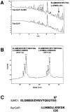
Identification of a linked peptide between RanGAP1 and SUMO-1. (A) Microbore C8 column profile of tryptic digests of SUMO-1–modified (top profile) and unmodified (bottom profile) RanGAP1, obtained by immunoprecipitation from solubilized rat liver NEs. Asterisks (*) indicate peaks unique to the SUMO-1–modified RanGAP1 that contain peptides derived from SUMO-1. Sequencing of the unique peak fraction eluting at ∼27 min (arrow) revealed the presence of two distinct sequences, one from SUMO-1 (ELGMEEDVIEVYXX...) and the other from RanGAP1 (LLIHGLLX...). (B) Matrix-assisted laser desorption time of flight (MALDI-TOF) mass spectrometric analysis of the 27-min peak reveals the presence of two peptides and their oxidation products (labeled with [O]) with mass values of 3.634 D (left peaks) and 3,877 D (right peaks). Based on the peptide sequences obtained in A and on the known protein sequences of RanGAP1 (Bischoff et al., 1995, DeGregori et al., 1994) and SUMO-1 (Mahajan et al., 1997), the observed mass values are consistent with two linked peptides involving the amino acid residues drawn above the peaks. (C) Depiction of the predicted isopeptide bond between the COOH-terminal glycine 97 of SUMO-1 and lysine 526 of RanGAP1.
Figure 2
Figure 2
SUMO-1 lacking the last four amino acids (SUMOΔC4) is a better substrate for modification of RanGAP1 in vitro than wt SUMO-1. Digitonin lysates of HeLa cells were mixed with RanGAP1 and ATP, and incubated for 10 min at RT in the absence (lane 2) or presence of wt SUMO-1 (lane 3), SUMOΔC4 (lane 4), GST–SUMO-1 (lane 5), or GST-SUMOΔC4 (lane 6). Lane 1 contains only HeLa extract. Efficiency of conversion of the 70-kD recombinant RanGAP1 (RanGAP) to a 90-kD SUMO-1–modified form (RanGAP•SUMO) or a ∼115-kD GST–SUMO-1–modified form (RanGAP•GST-SUMO) was assayed by immunoblotting with α-RanGAP1 antibodies.
Figure 3
Figure 3
SUMO-1 and SUMOΔC4, but not SUMOΔC6, modify multiple substrates in vivo. (A) Immunofluorescence of Cos-7 cells transfected with HA-tagged SUMO-1 constructs and probed with an α-HA monoclonal antibody. Intracellular localization of wt SUMO-1 (SUMO wt), SUMO-1 with a six–amino acid COOH-terminal deletion (SUMOΔC6), and SUMO-1 terminating at glycine 97 (SUMOΔC4) was analyzed. (B) Western blot analysis of SUMO-1–transfected cells. Cos-7 cells were transfected with HA-tagged wt SUMO-1 (lane 1), SUMOΔC6 (lane 2), or SUMOΔC4 (lane 3), and the cells were lysed in SDS gel loading buffer 24 h after transfection. Samples were electrophoresed on 8% (top) and 12.5% (bottom) polyacrylamide gels, transferred to nitrocellulose, and probed with the α-HA monoclonal antibody.
Figure 5
Figure 5
A single lysine residue (K526) in RanGAP1 is modified by SUMO-1. Bacterial lysates of cells expressing T7-tagged wt RanGAP1 (lanes 1 and 4) or mutant RanGAP1 (lanes 2 and 5) were mixed with a digitonin lysate of HeLa cells in the presence of ATP. After a 10-min incubation at RT the reaction products were analyzed by immunoblotting using mouse monoclonal antibodies against the T7 tag. Lanes 1 and 2, bacterial lysates before shift reaction; lanes 3–5, extracts after shift reaction. Lane 3, HeLa extract; lanes 4 and 5, HeLa extract and bacterial extract. The open arrowhead marks a proteolytic fragment of RanGAP1 that apparently is not competent for modification.
Figure 6
Figure 6
SUMO-1 modification of RanGAP1 at K526 is required for _targeting RanGAP1 to the nuclear rim in vivo. HA-tagged wt RanGAP1 (wt GAP) and mutant RanGAP1 (K526R GAP) were transfected into Cos-7 cells and detected after 24 h. (A) Localization of transfected HA-tagged proteins by indirect immunofluorescence. Cells were fixed and probed with an α-HA monoclonal antibody. (B) Western blot analysis of transfected cells. Transfected cells were lysed by scraping into boiling SDS gel loading buffer, and analyzed by immunoblotting with α-HA monoclonal antibody.
Figure 7
Figure 7
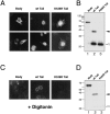
The mammalian-specific tail region of RanGAP1 is both necessary and sufficient for SUMO-1 modification and nuclear rim _targeting. (A) Immunofluorescence of Cos-7 cells transfected with HA-tagged RanGAP1 constructs representing the NH2-terminal 416 amino acids (Body), the COOH-terminal tail region containing amino acids 400-589 (wt Tail), and a mutant tail lacking the SUMO-1 modification site (K526R Tail). Transfected cells were fixed and probed with the α-HA monoclonal antibody. (B) Western blot analysis of transfected cells separated on a 12.5% polyacrylamide gel. Cells were transfected as above, lysed in boiling SDS gel loading buffer and analyzed by immunoblotting with α-HA monoclonal antibody. (C) Immunofluorescence of digitonin permeabilized Cos-7 cells transfected with the RanGAP1 constructs. Cells were permeabilized with 0.05% digitonin for 6 min, washed 3× in PBS before fixation, and then processed for immunofluorescence. (D) Immunoblots of digitonin-permeabilized transfected cells. Cells were permeabilized as described for immunofluorescence analysis, lysed in SDS gel loading buffer, and analyzed by immunoblotting with α-HA monoclonal antibodies. (B and D) Arrowheads indicate the positions of unmodified tail (open arrowhead) and modified tail (closed arrowhead).
Figure 8
Figure 8
Stable association of modified RanGAP1 with the NE during nuclear protein import. (A) Analysis of Cos-7 cells transfected with HA-tagged RanGAP1 by flow cytometry. Cos7 cells transfected with HA-RanGAP1 (A, top three panels), or mock-transfected cells (bottom panel) were permeabilized with digitonin and incubated in the presence of cytosol and ATP at the indicated temperature. To one sample recombinant RanGAP1 was added (30°C + RanGAP1). After 30 min, the cells were fixed and stained with α-HA monoclonal antibody and Cy5-conjugated secondary antibodies. The cell-associated Cy5 signal was detected by flow cytometry. (B) Analysis of nuclear import in Cos-7 cells transfected with HA-tagged RanGAP1. Cos7 cells transfected with HA-RanGAP1 were permeabilized with digitonin and incubated in the presence of cytosol, ATP, and FITC-labeled import substrate at the indicated temperature. To one sample recombinant RanGAP1 was added (30°C + RanGAP1). After 30 min, the cells were fixed, and processed for indirect immunofluorescence using α-HA monoclonal antibody. Arrows indicate corresponding cells in each set and are provided for orientation.
Figure 9
Figure 9
SUMO-1–mediated _targeting of RanGAP1 to the nuclear pore complex protein RanBP2. After SUMO-1 is proteolytically processed at the COOH terminus to expose glycine 97, it serves as the substrate in the ATP-dependent formation of an isopeptide bond between the free carboxyl group of glycine 97 in SUMO-1 and the ε-amino group of lysine 526 in the tail domain of RanGAP1. Modified RanGAP1 binds stably to RanBP2 at the cytoplasmic fibrils of the NPC through an interaction mediated by the SUMO-1–modified tail domain of RanGAP1. While the modification reaction is reversible, it remains to be seen whether RanGAP1 can be demodified after it has associated with RanBP2.

Similar articles

Cited by

References

    1. Adam SA, Sterne-Marr R, Gerace L. Nuclear protein import using digitonin-permeabilized cells. Methods Enzymol. 1992;219:97–110. - PubMed
    1. Arnason T, Ellison MJ. Stress resistance in Saccharomyces cerevisiaeis strongly correlated with assembly of a novel type of multiubiquitin chain. Mol Cell Biol. 1994;14:7876–7883. - PMC - PubMed
    1. Baboshina OV, Haas AL. Novel multiubiquitin chain linkages catalyzed by the conjugating enzymes E2EPF and RAD6 are recognized by 26 S proteasome subunit 5. J Biol Chem. 1996;271:2823–2831. - PubMed
    1. Baldi L, Brown K, Franzoso G, Siebenlist U. Critical role for lysines 21 and 22 in signal-induced, ubiquitin-mediated proteolysis of I κ B-α. J Biol Chem. 1996;271:376–379. - PubMed
    1. Biggins S, Ivanovska I, Rose MD. Yeast ubiquitin-like genes are involved in duplication of the microtubule organizing center. J Cell Biol. 1996;133:1331–1346. - PMC - PubMed

Publication types

  NODES
Association 7
twitter 2