. 1998 Jul;18(7):4262-71.
doi: 10.1128/MCB.18.7.4262.

Activated polo-like kinase Plx1 is required at multiple points during mitosis in Xenopus laevis

Affiliations

Activated polo-like kinase Plx1 is required at multiple points during mitosis in Xenopus laevis

Y W Qian et al. Mol Cell Biol. 1998 Jul.

Abstract

Entry into mitosis depends upon activation of the dual-specificity phosphatase Cdc25C, which dephosphorylates and activates the cyclin B-Cdc2 complex. Previous work has shown that the Xenopus polo-like kinase Plx1 can phosphorylate and activate Cdc25C in vitro. In the work presented here, we demonstrate that Plx1 is activated in vivo during oocyte maturation with the same kinetics as Cdc25C. Microinjection of wild-type Plx1 into Xenopus oocytes accelerated the rate of activation of Cdc25C and cyclin B-Cdc2. Conversely, microinjection of either an antibody against Plx1 or kinase-dead Plx1 significantly inhibited the activation of Cdc25C and cyclin B-Cdc2. This effect could be reversed by injection of active Cdc25C, indicating that Plx1 is upstream of Cdc25C. However, injection of Cdc25C, which directly activates cyclin B-Cdc2, also caused activation of Plx1, suggesting that a positive feedback loop exists in the Plx1 activation pathway. Other experiments show that injection of Plx1 antibody into early embryos, which do not require Cdc25C for the activation of cyclin B-Cdc2, resulted in an arrest of cleavage that was associated with monopolar spindles. These results demonstrate that in Xenopus laevis, Plx1 plays important roles both in the activation of Cdc25C at the initiation of mitosis and in spindle assembly at late stages of mitosis.


Figures
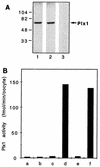
FIG. 1
FIG. 1
Characterization of affinity-purified Plx1 antibodies. (A) An extract of stage VI oocytes was prepared and subjected to immunoblotting. Lanes: 1 and 2, anti-Plx1 antibodies from two different rabbits; 3, control IgG. Molecular masses (in kilodaltons) are indicated on the left. (B) Immunoprecipitates were assayed for casein phosphorylation. Bars: a, c, and e, stage VI oocytes; b, d, and f, mature oocytes 15 min after undergoing GVBD; a and b, control IgG; c and d and e and f, anti-Plx1 antibodies from two different rabbits.
FIG. 2
FIG. 2
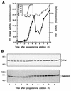
Activation of Plx1 during oocyte maturation. Oocytes were incubated in the presence of progesterone (3.2 μM), and groups of 10 oocytes were frozen at the times indicated. (A) Extracts were prepared, and histone H1 kinase (▪) and Plx1 (□) activities were determined. (Inset) The time of GVBD was established by examining oocytes for white spot formation with a dissecting microscope. (B) Samples of extracts were subjected to immunoblotting with anti-Plx1 or anti-Cdc25C antibodies (upper and lower panels, respectively). Molecular masses (in kilodaltons) are indicated on the left.
FIG. 3
FIG. 3
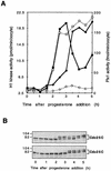
Delay of cyclin B-Cdc2 and Cdc25C activation by Plx1 antibodies. Oocytes were microinjected with anti-Plx1 or control IgG (75 ng per oocyte) and then, after 2 h, incubated in the presence of progesterone (3.2 μM). Groups of six oocytes were frozen at the times indicated. (A) Extracts were prepared, and histone H1 kinase and Plx1 activities were determined. Symbols for histone H1 kinase activity, ▪, control IgG; •, anti-Plx1 IgG. Symbols for Plx1 activity: □, control IgG; ○, anti-Plx1 IgG. (B) Samples of extracts were immunoblotted for Cdc25C. Upper panel, control IgG injection; lower panel, anti-Plx1 IgG injection. Molecular masses (in kilodaltons) are indicated on the left.
FIG. 4
FIG. 4
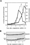
Reversal of the inhibitory effect of Plx1 antibodies by Plx1. All oocytes were microinjected with anti-Plx1 IgG (75 ng per oocyte) and then, after 2 h, with either recombinant Plx1 (60 ng per oocyte) or buffer. They were then incubated in the presence of progesterone (3.2 μM), and groups of six oocytes were frozen at the indicated times. (A) Extracts were prepared, and histone H1 kinase and Plx1 activities were determined. Symbols for histone H1 kinase activity: ▪, buffer injection; •, Plx1 injection; Symbols for Plx1 activity: □, buffer injection; ○, Plx1 injection. (B) Samples of extracts were immunoblotted for Cdc25C. Top, buffer injection; bottom, Plx1 injection. Molecular masses (in kilodaltons) are indicated on the left.
FIG. 5
FIG. 5
Reversal of the inhibitory effect of Plx1 antibodies by Cdc25C. All oocytes were microinjected with anti-Plx1 IgG (75 ng per oocyte) and then, after 2 h, with either wild-type Cdc25C or phosphatase-inactive Cdc25C(C457A) (25 ng per oocyte), and groups of six oocytes were frozen at the indicated times. Extracts were prepared, and histone H1 kinase and Plx1 activities were determined. Symbols for histone H1 kinase activity: ▪, Cdc25C(C457A); •, Cdc25C; Symbols for Plx1 activity: □, Cdc25C(C457A); ○, Cdc25C.
FIG. 6
FIG. 6
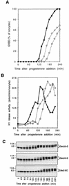
Acceleration of progesterone-induced maturation by Plx1. Oocytes were microinjected with either buffer, wild-type Plx1, or kinase-dead Plx1(N172A) (75 ng per oocyte) and then incubated in the presence of progesterone (48 nM). Groups of six oocytes were frozen at the indicated times. (A) Time course of maturation. Symbols: ▪, Plx1; ○, buffer; □, Plx1(N172A). (B) Extracts were prepared, and histone H1 kinase activity was determined. Symbols: ▪, Plx1; ○, buffer; □, Plx1(N172A). (C) Samples of extracts were immunoblotted for Cdc25C. Top, Plx1; middle, buffer; bottom, Plx1(N172A). Molecular masses (in kilodaltons) are indicated on the left.
FIG. 7
FIG. 7
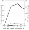
Activation of Plx1 in Cdc25C-injected oocytes. Oocytes were microinjected with either wild-type Cdc25C or phosphatase-inactive Cdc25C(C457A) (25 ng per oocyte), and groups of six oocytes were frozen at the indicated times. Extracts were prepared, and histone H1 kinase and Plx1 activities were determined. Symbols for histone H1 kinase activity: ▪, Cdc25C(C457A); •, Cdc25C. Symbols for Plx1 activity: □, Cdc25C(C457A); ○, Cdc25C.
FIG. 8
FIG. 8
Plx1 antibody arrests cleavage in early embryos. (A) Eggs were fertilized, and groups of 10 embryos were frozen at the indicated times. Extracts were prepared, and histone H1 kinase (▪) and Plx1 (□) activities were determined. (B) Plx1 antibody or control IgG was microinjected into one blastomere of a two-cell embryo, and embryonic development was monitored with a dissecting microscope. Plx1 antibody-injected blastomeres (right) ceased dividing one or two divisions after injection, whereas control antibody-injected cells continued to divide normally (left). (C) Cells in a late-blastula-stage embryo were fixed for confocal microscopy as described in Materials and Methods; a cell in early prophase is presented. a, Plx1 fluorescence; b, α-tubulin fluorescence of the same cell, staining spindle poles; c, merged image of a and b showing enrichment of Plx1 at the spindle poles. Bar, 15 μm. (D) Embryos were injected with either control IgG (a to c) or anti-Plx1 IgG (d to i) and processed for confocal microscopy as described in Materials and Methods. a, d, and g, DNA staining; b, e, and h, α-tubulin fluorescence staining; c, f, and i, merged images of a and b, d and e, and g and h, respectively. Bars, 15 μm.
FIG. 8
FIG. 8
Plx1 antibody arrests cleavage in early embryos. (A) Eggs were fertilized, and groups of 10 embryos were frozen at the indicated times. Extracts were prepared, and histone H1 kinase (▪) and Plx1 (□) activities were determined. (B) Plx1 antibody or control IgG was microinjected into one blastomere of a two-cell embryo, and embryonic development was monitored with a dissecting microscope. Plx1 antibody-injected blastomeres (right) ceased dividing one or two divisions after injection, whereas control antibody-injected cells continued to divide normally (left). (C) Cells in a late-blastula-stage embryo were fixed for confocal microscopy as described in Materials and Methods; a cell in early prophase is presented. a, Plx1 fluorescence; b, α-tubulin fluorescence of the same cell, staining spindle poles; c, merged image of a and b showing enrichment of Plx1 at the spindle poles. Bar, 15 μm. (D) Embryos were injected with either control IgG (a to c) or anti-Plx1 IgG (d to i) and processed for confocal microscopy as described in Materials and Methods. a, d, and g, DNA staining; b, e, and h, α-tubulin fluorescence staining; c, f, and i, merged images of a and b, d and e, and g and h, respectively. Bars, 15 μm.

Similar articles

Cited by

References

    1. Amon A, Surana U, Muroff I, Nasmyth K. Regulation of p34CDC28 tyrosine phosphorylation is not required for entry into mitosis in S. cerevisiae. Nature. 1992;355:368–371. - PubMed
    1. Dasso M, Newport J W. Completion of DNA replication is monitored by a feedback system that controls the initiation of mitosis in vitro: studies in Xenopus. Cell. 1990;61:811–823. - PubMed
    1. Davis F M, Tsao T Y, Fowler S K, Rao P N. Monoclonal antibodies to mitotic cells. Proc Natl Acad Sci USA. 1983;80:2926–2930. - PMC - PubMed
    1. Dunphy W G, Kumagai A. The cdc25 protein contains an intrinsic phosphatase activity. Cell. 1991;67:189–196. - PubMed
    1. Edgar B A, Lehman D A, O’Farrell P H. Transcriptional regulation of string (cdc25): a link between developmental programming and the cell cycle. Development. 1994;120:3131–3143. - PMC - PubMed

Publication types

  NODES
twitter 2