Comparative Study
. 1999 Feb;19(2):1092-100.
doi: 10.1128/MCB.19.2.1092.

Two polymorphic variants of wild-type p53 differ biochemically and biologically

Affiliations
Comparative Study

Two polymorphic variants of wild-type p53 differ biochemically and biologically

M Thomas et al. Mol Cell Biol. 1999 Feb.

Abstract

The wild-type p53 protein exhibits a common polymorphism at amino acid 72, resulting in either a proline residue (p53Pro) or an arginine residue (p53Arg) at this position. Despite the difference that this change makes in the primary structure of the protein resulting in a difference in migration during sodium dodecyl sulfate-polyacrylamide gel electrophoresis, no differences in the biochemical or biological characteristics of these wild-type p53 variants have been reported. We have recently shown that p53Arg is significantly more susceptible than p53Pro to the degradation induced by human papillomavirus (HPV) E6 protein. Moreover, this may result in an increased susceptibility to HPV-induced tumors in homozygous p53Arg individuals. In further investigating the characteristics of these p53 variants, we now show that both forms are morphologically wild type and do not differ in their ability to bind to DNA in a sequence-specific manner. However, there are a number of differences between the p53 variants in their abilities to bind components of the transcriptional machinery, to activate transcription, to induce apoptosis, and to repress the transformation of primary cells. These observations may have implications for the development of cancers which harbor wild-type p53 sequences and possibly for the ability of such tumors to respond to therapy, depending on their p53 genotype.


Figures

FIG. 1
FIG. 1
p53Pro and p53Arg display similar epitopes. Immunoprecipitation analysis of in vitro-translated p53Pro and p53Arg proteins with a panel of antibodies is shown. The variants reacted equally well with the wild-type-specific PAb1620 antibody and with the carboxy-terminal-specific C4 and PAb421 antibodies. No reaction is observed with the mutant-specific PAb240 antibody or with the murine-specific PAb246 antibody. The right-hand panel shows the level of input proteins.
FIG. 2
FIG. 2
(A) Comparison of p53Pro and p53Arg transcriptional activation of pCONCAT and pG13CAT. The upper panels show the transcriptional activities of the wild-type p53 variants and mutant p53 protein determined by using two different reporter CAT plasmids, p53CONCAT and pG13CAT. p53-null 10(1) murine fibroblast cells were cotransfected with p53 expression plasmids or a control empty plasmid and the CAT reporter plasmid, and the CAT activity in the transfected cells was determined. p53Arg and p53Pro represent the two wild-type variants of p53, and Mp53-175 is the inactive p53 point mutant. The percent CAT conversion is also shown. The lower panels show Western blot analysis of the same cell lysates used in the CAT assay and demonstrate that equal amounts of plasmid-derived p53 protein were present in the transfected cells. (B) Comparison of p53Pro and p53Arg transcriptional activation of the of the p21 promoter by using a luciferase assay. The upper panel shows transcriptional activity of the wild-type p53 variants and mutant p53 protein determined by using the luciferase reporter plasmid pWT3L2, which contains the p53-responsive p21 promoter and enhancer. The lower panel shows Western blot analysis of the same cell lysates used in the luciferase assays and demonstrates that equal amounts of plasmid-derived p53 protein were present in the transfected cell lysates.
FIG. 3
FIG. 3
Comparison of p53Pro and p53Arg transcriptional activation following transfection of various concentrations of p53 variant-expressing plasmids. Between 1.0 and 0.0025 μg of p53Arg- or p53Pro-expressing plasmid was cotransfected with 5.0 μg of p53CONCAT plasmid as indicated. As a negative control for p53 activity, the p53CONCAT plasmid was cotransfected with 3 μg of the Mp53-175 plasmid. The percent CAT conversion is shown.
FIG. 4
FIG. 4
Comparison of sequence-specific DNA binding activities of p53Pro and p53Arg. (A) Comparison of the abilities of p53Arg and p53Pro to bind specific enhancer sequences (bax, CON*, and p53CON). p53 proteins and control HPV-18 E6 protein were synthesized in vitro and preincubated in the presence (+) or absence (−) of the monoclonal antibody PAb421, which activates p53 sequence-specific binding. Proteins were incubated in the presence of the labeled DNA sequence at room temperature for 40 min and then run on a nondenaturing gel. Note that the higher-molecular-weight specific binding band was obtained for the p53 variants only in the presence of PAb421, and no specific binding was obtained with the E6 protein. As shown at the bottom, a portion of the synthesized p53 proteins was also labeled and run on an SDS-polyacrylamide gel in order to ensure that the same amounts of p53 protein were synthesized and assayed in each reaction. (B) Titration of p53Pro and p53Arg binding to p53CON DNA. Decreasing amounts of unlabeled p53Arg and p53Pro proteins synthesized in vitro were preincubated in the presence of the monoclonal antibody PAb421 and then incubated in the presence of the labeled p53CON DNA sequence at room temperature for 40 min and run on a nondenaturing gel. As shown on the bottom, a portion of the synthesized p53 proteins was also labeled and run on an SDS-polyacrylamide gel in order to ensure that the same amounts of p53 protein were synthesized and assayed in each reaction.
FIG. 5
FIG. 5
Interactions of p53Pro and p53Arg with transcription factors. (A) GST fusion protein pull-down assay with GST-p53Pro and GST-p53Arg bound to glutathione-agarose. In vitro-translated (ivt) 35S-labeled p53Pro, p53Arg, TAFII70, or TBP proteins were incubated with the fusion protein-agarose at either 0°C or room temperature (rt). The bound proteins were analyzed by SDS-PAGE and autoradiography. No differences in the abilities of p53Pro and p53Arg to form either hetero- or homodimers or in their abilities to bind to TBP were apparent. However, TAFII70 is bound significantly only at room temperature, and p53Pro binds more strongly than p53Arg to TAFII70. (B) The GST-fusion protein pull-down assay repeated with in vitro-translated 35S-labeled TAFII32 and TAFII250 plus p53Pro (P) and p53Arg (R) for comparison. TAFII32 binds more strongly at room temperature and binds more strongly to p53Pro than to p53Arg. TAFII250 binds to p53Pro and p53Arg with similar affinities but binds more strongly at room temperature. (C) Phosphorimager analysis of at least five GST fusion protein pull-down assays. The fold increase in binding at room temperature over binding at 0°C is shown. p53Pro binds approximately twice as strongly as p53Arg to TAFII32 and TAFII70. GST alone was also used as a negative control; in all cases, the percentage of loaded protein retained was less than 1%.
FIG. 5
FIG. 5
Interactions of p53Pro and p53Arg with transcription factors. (A) GST fusion protein pull-down assay with GST-p53Pro and GST-p53Arg bound to glutathione-agarose. In vitro-translated (ivt) 35S-labeled p53Pro, p53Arg, TAFII70, or TBP proteins were incubated with the fusion protein-agarose at either 0°C or room temperature (rt). The bound proteins were analyzed by SDS-PAGE and autoradiography. No differences in the abilities of p53Pro and p53Arg to form either hetero- or homodimers or in their abilities to bind to TBP were apparent. However, TAFII70 is bound significantly only at room temperature, and p53Pro binds more strongly than p53Arg to TAFII70. (B) The GST-fusion protein pull-down assay repeated with in vitro-translated 35S-labeled TAFII32 and TAFII250 plus p53Pro (P) and p53Arg (R) for comparison. TAFII32 binds more strongly at room temperature and binds more strongly to p53Pro than to p53Arg. TAFII250 binds to p53Pro and p53Arg with similar affinities but binds more strongly at room temperature. (C) Phosphorimager analysis of at least five GST fusion protein pull-down assays. The fold increase in binding at room temperature over binding at 0°C is shown. p53Pro binds approximately twice as strongly as p53Arg to TAFII32 and TAFII70. GST alone was also used as a negative control; in all cases, the percentage of loaded protein retained was less than 1%.
FIG. 6
FIG. 6
p53 induction pathways are functional in stably transfected BRK cell lines. Western blot analysis of the p53 protein in pools of colonies obtained after cotransfection of BRK cells with either p53Pro or p53Arg, with or without UV irradiation, is shown.
FIG. 7
FIG. 7
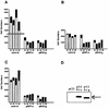
Comparison of the activities of p53Pro and p53Arg in inducing apoptosis. (A, B, and C) Saos-2 cells were transfected with either pCDNA3 (control vector), pCDp53Pro, or pCDp53Arg together with a lacZ-expressing plasmid. Cells were fixed at the times posttransfection indicated and stained for lacZ expression. Histograms show the number of live (white bars) and dead (black bars) blue cells present at 24, 48, 72, and 96 h posttransfection. Each panel shows an independent assay. (D) Western blot analysis of a parallel transfection harvested at 24 h confirms equivalent levels of p53Pro and p53Arg expression.

Similar articles

Cited by

References

    1. Banks L, Matlashewski G, Crawford L. Isolation of human-p53-specific monoclonal antibodies and their use in the studies of human p53 expression. Eur J Biochem. 1986;159:529–534. - PubMed
    1. Beckman G, Birgander R, Sjalander A, Saha N, Holmberg P, Kiveld S, Beckman L. Is p53 polymorphism maintained by natural selection. Hum Hered. 1994;44:266–270. - PubMed
    1. Caelles C, Helmberg A, Karin M. p53-dependent apoptosis in the absence of transcriptional activation of p53-_target genes. Nature. 1994;370:220–223. - PubMed
    1. Cho Y, Gorina S, Jeffrey P, Pavletich N. Crystal structure of a p53 tumour suppressor-DNA complex: understanding tumorigenic mutations. Science. 1994;265:346–355. - PubMed
    1. Donehower L, Harvey B, Slagle B, McArthur B, Montgomery C, Butel J, Bradley A. Mice deficient for p53 are developmentally normal but susceptible to spontaneous tumours. Nature. 1992;356:215–221. - PubMed

Publication types

MeSH terms

  NODES
INTERN 2
Note 1
twitter 2